<< Chapter < Page Chapter >> Page >
a) a micrograph of a large oval (10 µm) labeled ascus and smaller ovals (5 µm) labeled ascospores. B) a micrograph of a long stalk with strands of spheres emanating from a sphere on the tip. The spheres are about 2 µm in diameter. C) A long strand with clusters of spheres. A small dot in each sphere is labeled nucleus.
(a) This brightfield micrograph shows ascospores being released from asci in the fungus Talaromyces flavus var. flavus . (b) This electron micrograph shows the conidia (spores) borne on the conidiophore of Aspergillus , a type of toxic fungus found mostly in soil and plants. (c) This brightfield micrograph shows the yeast Candida albicans, the causative agent of candidiasis and thrush. (credit a, b, c: modification of work by Centers for Disease Control and Prevention)
A micrograph showing a thick tube with 8 ovals lined up within the tube.
These ascospores, lined up within an ascus, are produced sexually. (credit: Peter G. Werner)
Ascomycete life cycle. Mycelia produce conidiophores which use mitosis to asexually produce spores. These spores then germinate into new mycelia. Sexual reproduction begins one hyphae produces an ascogonium and another produces an antheridium. In plasmogamy the ascogonium and antheridium fuse. Mitosis and cell division result in the formation of many dikaryotic hyphae, which form a fruiting body called the ascocarp. Asci form at the tips of these hyphae. In karyogamy the nuclein in the asci fuse to form a diploid zygote. Then meiosis produces four haploid nuclei in the ascus. Then mitosis and cell division results in eight haploid ascospores in the ascus. These ascospores then disperse and germinate into new mycelia.
The life cycle of an ascomycete is characterized by the production of asci during the sexual phase. The haploid phase is the predominant phase of the life cycle.

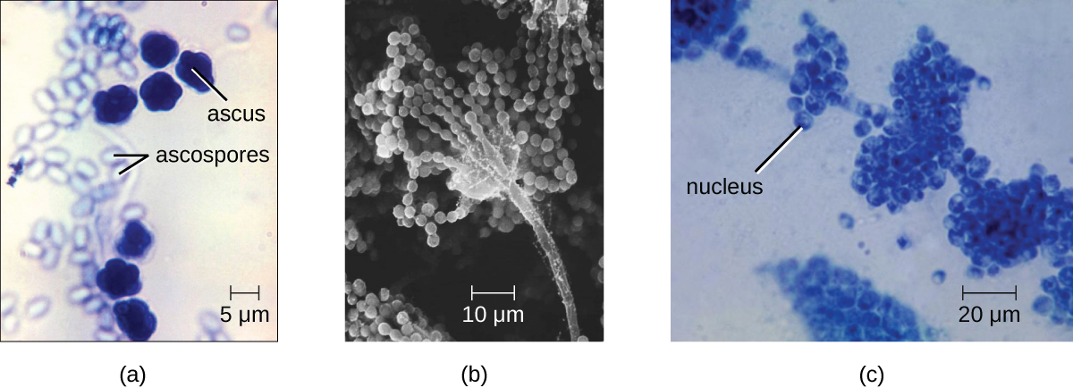
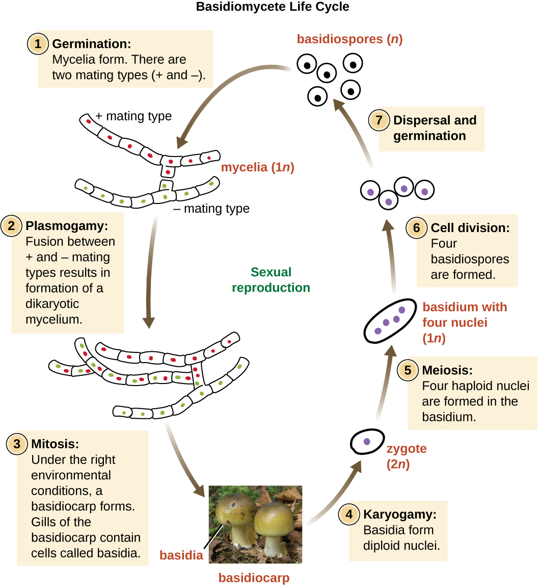
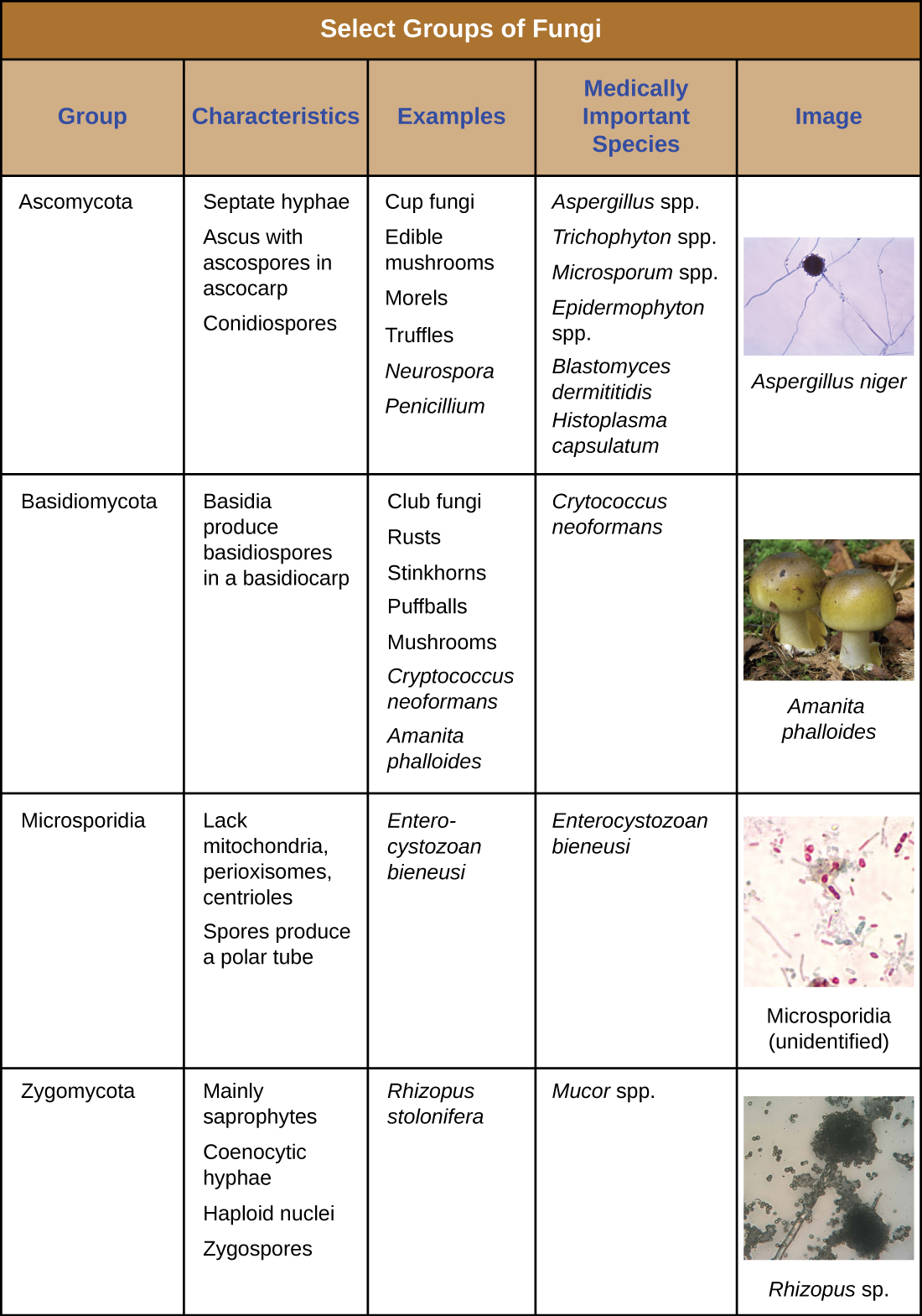
The Basidiomycota (basidiomycetes) are fungi that have basidia (club-shaped structures) that produce basidiospores (spores produced through budding) within fruiting bodies called basidiocarps ( [link] ). They are important as decomposers and as food. This group includes rusts, stinkhorns, puffballs, and mushrooms. Several species are of particular importance. Cryptococcus neoformans , a fungus commonly found as a yeast in the environment, can cause serious lung infections when inhaled by individuals with weakened immune systems. The edible meadow mushroom, Agricus campestris , is a basidiomycete, as is the poisonous mushroom Amanita phalloides , known as the death cap. The deadly toxins produced by A. phalloides have been used to study transcription.

Basidiomycete life cycle. Haploid basidiospres germinate to form mycelia. There are two mating types (+ and -_). In plasmogamy, fusion between + and – mating types results in formation of a dikaryotic mycelium. Under the right environmental conditions, a basidiocarp forms via mitosis. Gills of the basidiocarp contain cells called basidia. A photo of a mushroom labels the mushroom as basidiocap and basidia within the gills. Basidia form diploid nuclei via karyotamy; this produces a diploid zygote. Four haploid nuclei are formed in the basidium via meisos. Cell division produces four haploid basidiospores. These spres then disperse and germinate into new mycelia.
The life cycle of a basidiomycete alternates a haploid generation with a prolonged stage in which two nuclei (dikaryon) are present in the hyphae.

Finally, the Microsporidia are unicellular fungi that are obligate intracellular parasites. They lack mitochondria, peroxisomes, and centrioles, but their spores release a unique polar tubule that pierces the host cell membrane to allow the fungus to gain entry into the cell. A number of microsporidia are human pathogens, and infections with microsporidia are called microsporidiosis . One pathogenic species is Enterocystozoan bieneusi , which can cause symptoms such as diarrhea, cholecystitis (inflammation of the gall bladder), and in rare cases, respiratory illness.

A table labeled select groups of fungi. Four groups are discussed. Ascomycota have the characteristics: septate hyphae, ascus with ascospores in ascocarp, and conidiospores. Examples include Cup fungi, edible mushrooms, morels, truffles, neurospora, and penicillim. Medically important species include Aspergillus, Trichophyton, Microsporum, Epidemophyton, Blastomyces demititidis, and Histoplasma capsulatum. An image of Aspergillus niger shows long strands with a dark sphere at the end of one strand. Basidiomycota have the characteristics: basidia, produce basidiospores in basidiocarp. Examples include club fungi, rusts, stinkhors, puffballs, mushrooms, Cryptococcus neoformans, Amanita phalloides. Medically important species include Cryptococcus neoformans. An image shows a mushroom labeled Amanita phalloides. Microsporidia have the characteristics: lack mitochondria, peroxisomes, and centrioles; spores produce a polar tube. Examples include Enterocystozoan bieneusi which is medically important. A micrograph shows oval cells labeled microsporidia (unidentified). Zygomycota have the characteristics: mainly saprophytes, coenocytic hyphae, haploid nuclei and zygospores. Examples include Rhizopus stolonifera and the medically important mucor spp. A micrograph shows a long strand with many small dots everywhere on the slide.
(credit “Ascomycota”: modification of work by Dr. Lucille Georg, Centers for Disease Control and Prevention; credit “Microsporidia”: modification of work by Centers for Disease Control and Prevention)
  • Which group of fungi appears to be associated with the greatest number of human diseases?

Eukaryotic pathogens in eukaryotic hosts

When we think about antimicrobial medications, antibiotics such as penicillin often come to mind. Penicillin and related antibiotics interfere with the synthesis of peptidoglycan cell walls, which effectively targets bacterial cells. These antibiotics are useful because humans (like all eukaryotes) do not have peptidoglycan cell walls.

Developing medications that are effective against eukaryotic cells but not harmful to human cells is more difficult. Despite huge morphological differences, the cells of humans, fungi, and protists are similar in terms of their ribosomes, cytoskeletons, and cell membranes. As a result, it is more challenging to develop medications that target protozoans and fungi in the same way that antibiotics target prokaryotes.

Fungicides have relatively limited modes of action. Because fungi have ergosterols (instead of cholesterol) in their cell membranes, the different enzymes involved in sterol production can be a target of some medications. The azole and morpholine fungicides interfere with the synthesis of membrane sterols. These are used widely in agriculture (fenpropimorph) and clinically (e.g., miconazole). Some antifungal medications target the chitin cell walls of fungi. Despite the success of these compounds in targeting fungi, antifungal medications for systemic infections still tend to have more toxic side effects than antibiotics for bacteria.

Part 3

Sarah is relieved the ringworm is not an actual worm, but wants to know what it really is. The physician explains that ringworm is a fungus. He tells her that she will not see mushrooms popping out of her skin, because this fungus is more like the invisible part of a mushroom that hides in the soil. He reassures her that they are going to get the fungus out of her too.

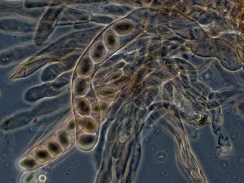
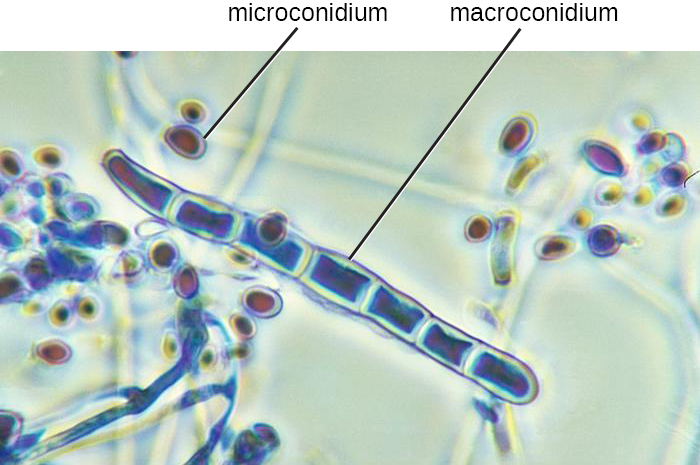
The doctor cleans and then carefully scrapes the lesion to place a specimen on a slide. By looking at it under a microscope, the physician is able to confirm that a fungal infection is responsible for Sarah’s lesion. In [link] , it is possible to see macro- and microconidia in Trichophyton rubrum . Cell walls are also visible. Even if the pathogen resembled a helminth under the microscope, the presence of cell walls would rule out the possibility because animal cells lack cell walls.

The doctor prescribes an antifungal cream for Sarah’s mother to apply to the ringworm. Sarah’s mother asks, “What should we do if it doesn’t go away?”

  • Can all forms of ringworm be treated with the same antifungal medication?
A micrograph of a long strands with cell walls. The long strand is labeled macroconidium. Smaller spheres outside the long strand are labeled microconidia.
This micrograph shows hyphae (macroconidium) and microconidia of Trichophyton rubrum , a dermatophyte responsible for fungal infections of the skin. (credit: modification of work by Centers for Disease Control and Prevention)

Jump to the next Clinical Focus box. Go back to the previous Clinical Focus box.

Key concepts and summary

  • The fungi include diverse saprotrophic eukaryotic organisms with chitin cell walls
  • Fungi can be unicellular or multicellular; some (like yeast) and fungal spores are microscopic, whereas some are large and conspicuous
  • Reproductive types are important in distinguishing fungal groups
  • Medically important species exist in the four fungal groups Zygomycota, Ascomycota, Basidiomycota, and Microsporidia
  • Members of Zygomycota, Ascomycota, and Basidiomycota produce deadly toxins
  • Important differences in fungal cells, such as ergosterols in fungal membranes, can be targets for antifungal medications, but similarities between human and fungal cells make it difficult to find targets for medications and these medications often have toxic adverse effects

Fill in the blank

Nonseptate hyphae are also called _________.

coenocytic

Got questions? Get instant answers now!

Unicellular fungi are called _________.

yeasts

Got questions? Get instant answers now!

Some fungi have proven medically useful because they can be used to produce _________.

antibiotics

Got questions? Get instant answers now!

Short answer

Which genera of fungi are common dermatophytes (fungi that cause skin infections)?

Got questions? Get instant answers now!

What is a dikaryotic cell?

Got questions? Get instant answers now!

Get Jobilize Job Search Mobile App in your pocket Now!

Get it on Google Play Download on the App Store Now


Source:  OpenStax, Microbiology. OpenStax CNX. Nov 01, 2016 Download for free at http://cnx.org/content/col12087/1.4
Google Play and the Google Play logo are trademarks of Google Inc.

Notification Switch

Would you like to follow the 'Microbiology' conversation and receive update notifications?

Ask