

| << Chapter < Page | Chapter >> Page > |
Figure
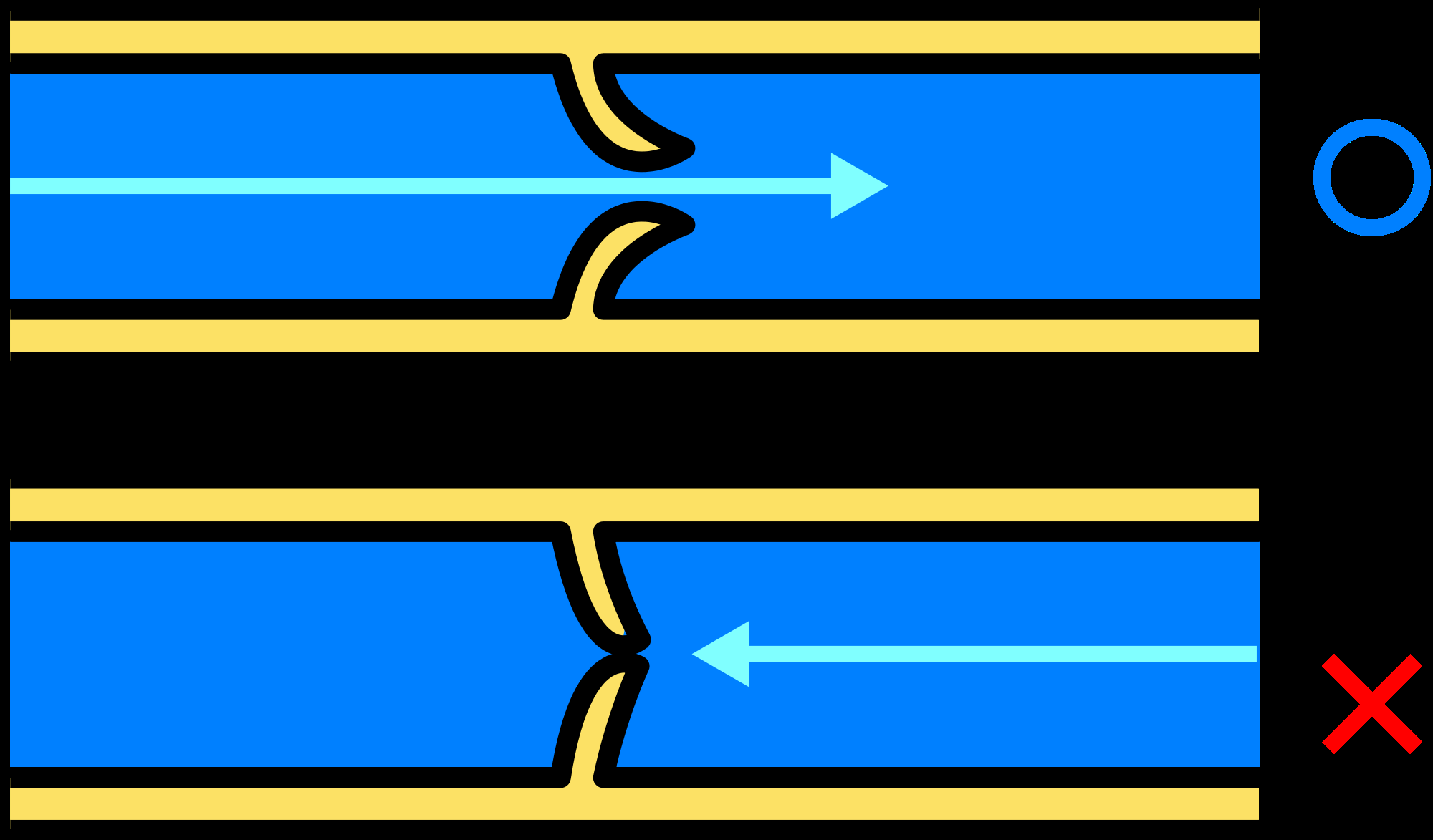
Shows the how valves cause blood to only flow one way though veins
http://upload.wikimedia.org/wikipedia/commons/thumb/4/4a/Venous_valve.svg /2000px-Venous_valve.svg.png
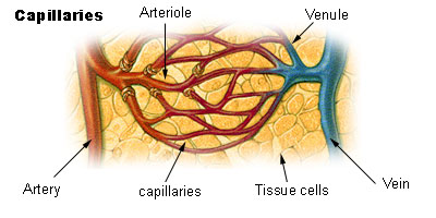
Figure showing capillaries as the transition between arteries and veins
http://en.wikipedia.org/wiki/File:Illu_capillary.jpg
Interactive diagram illustrating arterial and venous structure.
http://www.phschool.com/science/biology_place/biocoach/cardio2/structure.html
Figure
Shows the how valves cause blood to only flow one way though veins
http://upload.wikimedia.org/wikipedia/commons/thumb/4/4a/Venous_valve.svg /2000px-Venous_valve.svg.png
| Artery | Vein |
| Small lumen | Large lumen |
| Blood under high pressure | Blood under low pressure |
| Valves absent | Valves present |
| Carries blood away from heart | Carries blood towards the heart |
| Carries oxygenated blood except pulmonary artery | Carries deoxygenated blood except pulmonary vein |
Use and symbology of blood and heart in traditional black culture
Investigation: Practical investigation of sheep’s heart
Video: Doing a dissection
Equipment:
|
|
1. EXTERNAL
(a)How would you describe the general shape of the heart?
(b)Note the grooves on the surface of the heart. In which direction do they run.
What do you observe in these grooves.
(c)Identify the atria and ventricles. How do they differ from each other in
appearance. What difference do you notice between the atria and ventricles.
2. If the venae cavae are sufficiently long, insert a funnel into the superior vena cava and tie off the inferior vena cava with a piece of cotton . When water is added through the superior vena cave into the right atrium:
(a)What happens to the wall of the right ventricle?
(b)Press the right ventricle. What do you observe?
(c)Release the pressure. What happens?
(d)Now press the left ventricle a few times. What do you notice?
(e)Now attach funnel to one of the pulmonary veins and tie off the others
(if possible). Pour water down the funnel and press the left ventricle.
What do you observe?
(f)Release the pressure and press the right ventricle. What do you observe?
Remove the funnel and tubes.
3. Cut the superior vena cava from the atrium and cut open the wall of the atrium. Dothe same with the pulmonary vein and left atrium.
(a)Describe the appearance of the inner atrial surface.
(b)Determine the position of the pulmonary artery and the aorta by inserting a
glass rod through these vessel into the chambers of the heart.
Name the artery that leaves the right ventricle.
Name the artery that leaves the left ventricle.
4. Make an incision in the right side of the left ventricle from the oblique groove to the a pex of the heart.
(a)What do you observe between the left atrium and left ventricle?
(b)How many flaps do you see?
(c)What is the function of these flaps?
5. Similarly, make an incision in the left wall of the right ventricle from the oblique groove.
(a)How many flaps do you see between the atrium and the ventricle?
(b)What do these flaps collectively form?
6. Compare the muscular walls of the:
(a) atria and the ventricles
(b) left and right ventricles
7. What do you observe between the two halves of the heart.
8. Examine the tendinous cords .
(a)Where are their points of attachment?
(b)What is their function
9. If the pulmonary artery and aorta are long enough, do this question. Using a funnel, pour water into the pulmonary artery and the aorta.
(a)What do you notice?
(b)What do you see at the base of these arteries?
10. Cut the aorta and pulmonary arteries open longitudinally and examine the valves.
(a)How many parts are there to each of these valves?
(b)Compare the walls of the aorta and the pulmonary artery and suggest a
reason for any difference you many find.
Notification Switch
Would you like to follow the 'Siyavula: life sciences grade 10' conversation and receive update notifications?
|
|

|
|

|
|

|
|

|
|

|
|
|
|

|
|
|
|
|
|
|