Figure: Simplified Diagrammatic sketch of the entire circulatory system. Blood
flows to every inch of the body, even to the tips of the fingers and toes.Lungs provide oxygen to the blood. The digestive system supplies nutrients. The
kidneys filter the blood.
The heart and associated blood vessels
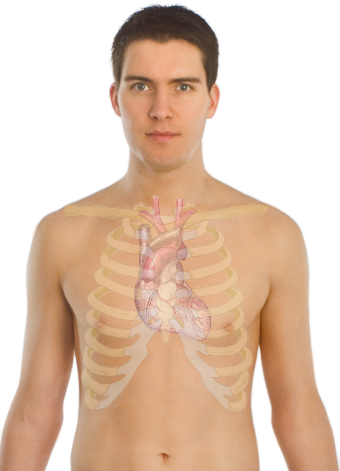
Figure 1 : Heart overlayed on a body so show the location of the heart within
the chest.
(External Link)
The heart is situated in your thorax just behind your breastbone and is about
the size of your fist.
It is a large muscle that pumps through repeated rhythmic contractions and
therefore requires lots of nutrients and oxygen.
On the surface of the heart are
coronary arteries that are arteries that branch off the aorta and supply the heart with oxygen and
nutrients.
The heart is made up of 4 chambers and divided by a
septum into a right and left half.
The right half of the heart pumps
deoxygenated blood up into the pulmonary artery, towards the lungs (pulmonary circulation),
where it is oxygenated.
Oxygenated blood returns from the lungs via the pulmonary veins and enters the
left side of the heart.
The left side of the heart then pumps
oxygenated blood up through the aorta, and into the general circulation (systemic
circulation) and the oxygen is consumed by the body.
Deoxygenated blood returns to the right side of the heart via the inferior vena
cava which drains blood from below the heart and superior vena cava, whichbrings blood from the head and arms.
The human circulatory system is a double circulatory system, because blood
travels to the heart twice during circulation, once before going to the lungsand once before circulating throughout the body.
Blood only flows in one direction, through the circulatory system.
All vessels that flow
A way from the heart are called
A rteries.
All blood vessels entering the heart are called
V eins.
The terms artery and vein are
not
determined by what the vessel transports (oxygenated blood or deoxygenated) but
by whether the vessel flows to or from the heart.
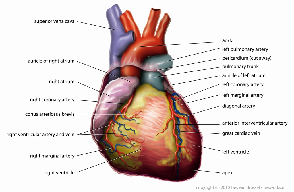
Figure 2 : General structure of the heart and associated blood vessels
(http://en.wikipedia.org/wiki/File:Anatomy_Heart_English_Tiesworks.jpg)
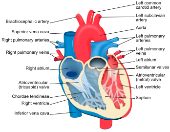
Internal structure of the heart
The heart is made up of
4 chambers. There are
2 atria at the top of the heart which receives blood and
2 ventricles at the bottom of the heart which pumps blood out of the heart.
The
septum divides the left and right side of the heart.
The valves of the heart ensure that blood only flows one way through the heart.
The
tricuspid valve is found between the right atrium and the right ventricle.
The
mitral valve is found between the left atrium and the left ventricle.
Strong tendinous chords attached to valves prevent them from turning inside out
when they close.
The
semi-lunar valves are located at the bottom of the aorta and pulmonary artery.
Internal structure of the heart
Interesting facts : Humans, birds, and mammals have a four-chambered heart. Fish have a two-
chambered heart, one atrium and one ventricle. Amphibians have a three-chambered heart with two atria and one ventricle. The advantage of a four
chambered heart is that there is no mixture of the oxygenated and deoxygenatedblood.