<< Chapter < Page Chapter >> Page >

The mitotic phase

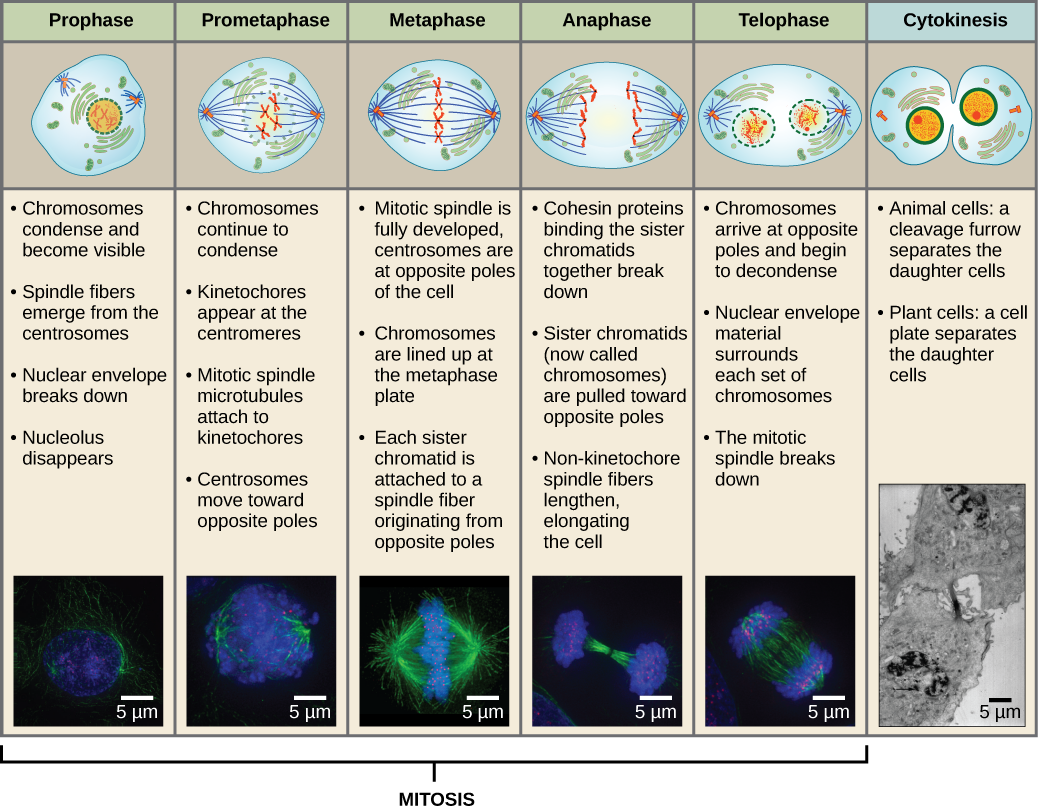
The mitotic phase is a multistep process during which the duplicated chromosomes are aligned, separated, and move into two new, identical daughter cells. The first portion of the mitotic phase is called karyokinesis    , or nuclear division. The second portion of the mitotic phase, called cytokinesis, is the physical separation of the cytoplasmic components into the two daughter cells.

Revisit the stages of mitosis at this site .

Karyokinesis (mitosis)

Karyokinesis, also known as mitosis    , is divided into a series of phases—prophase, prometaphase, metaphase, anaphase, and telophase—that result in the division of the cell nucleus ( [link] ). Karyokinesis is also called mitosis.

Art connection

This diagram shows the five phases of mitosis and cytokinesis. During prophase, the chromosomes condense and become visible, spindle fibers emerge from the centrosomes, the nuclear envelope breaks down, and the nucleolus disappears. During prometaphase, the chromosomes continue to condense and kinetochores appear at the centromeres. Mitotic spindle microtubules attach to the kinetochores, and centrosomes move toward opposite poles. During metaphase, the mitotic spindle is fully developed, and centrosomes are at opposite poles of the cell. Chromosomes line up at the metaphase plate and each sister chromatid is attached to a spindle fiber originating from the opposite pole. During anaphase, the cohesin proteins that were binding the sister chromatids together break down. The sister chromatids, which are now called chromosomes, move toward opposite poles of the cell. Non-kinetochore spindle fibers lengthen, elongating the cell. During telophase, chromosomes arrive at the opposite poles and begin to decondense. The nuclear envelope reforms. During cytokinesis in animals, a cleavage furrow separates the two daughter cells. In plants, a cell plate separates the two cells.
Karyokinesis (or mitosis) is divided into five stages—prophase, prometaphase, metaphase, anaphase, and telophase. The pictures at the bottom were taken by fluorescence microscopy (hence, the black background) of cells artificially stained by fluorescent dyes: blue fluorescence indicates DNA (chromosomes) and green fluorescence indicates microtubules (spindle apparatus). (credit “mitosis drawings”: modification of work by Mariana Ruiz Villareal; credit “micrographs”: modification of work by Roy van Heesbeen; credit “cytokinesis micrograph”: Wadsworth Center/New York State Department of Health; scale-bar data from Matt Russell)

Which of the following is the correct order of events in mitosis?

  1. Sister chromatids line up at the metaphase plate. The kinetochore becomes attached to the mitotic spindle. The nucleus reforms and the cell divides. Cohesin proteins break down and the sister chromatids separate.
  2. The kinetochore becomes attached to the mitotic spindle. Cohesin proteins break down and the sister chromatids separate. Sister chromatids line up at the metaphase plate. The nucleus reforms and the cell divides.
  3. The kinetochore becomes attached to the cohesin proteins. Sister chromatids line up at the metaphase plate. The kinetochore breaks down and the sister chromatids separate. The nucleus reforms and the cell divides.
  4. The kinetochore becomes attached to the mitotic spindle. Sister chromatids line up at the metaphase plate. Cohesin proteins break down and the sister chromatids separate. The nucleus reforms and the cell divides.

During prophase    , the “first phase,” the nuclear envelope starts to dissociate into small vesicles, and the membranous organelles (such as the Golgi complex or Golgi apparatus, and endoplasmic reticulum), fragment and disperse toward the periphery of the cell. The nucleolus disappears (disperses). The centrosomes begin to move to opposite poles of the cell. Microtubules that will form the mitotic spindle extend between the centrosomes, pushing them farther apart as the microtubule fibers lengthen. The sister chromatids begin to coil more tightly with the aid of condensin    proteins and become visible under a light microscope.

During prometaphase    , the “first change phase,” many processes that were begun in prophase continue to advance. The remnants of the nuclear envelope fragment. The mitotic spindle continues to develop as more microtubules assemble and stretch across the length of the former nuclear area. Chromosomes become more condensed and discrete. Each sister chromatid develops a protein structure called a kinetochore    in the centromeric region ( [link] ). The proteins of the kinetochore attract and bind mitotic spindle microtubules. As the spindle microtubules extend from the centrosomes, some of these microtubules come into contact with and firmly bind to the kinetochores. Once a mitotic fiber attaches to a chromosome, the chromosome will be oriented until the kinetochores of sister chromatids face the opposite poles. Eventually, all the sister chromatids will be attached via their kinetochores to microtubules from opposing poles. Spindle microtubules that do not engage the chromosomes are called polar microtubules. These microtubules overlap each other midway between the two poles and contribute to cell elongation. Astral microtubules are located near the poles, aid in spindle orientation, and are required for the regulation of mitosis.

Questions & Answers

calculate molarity of NaOH solution when 25.0ml of NaOH titrated with 27.2ml of 0.2m H2SO4
Gasin Reply
what's Thermochemistry
rhoda Reply
the study of the heat energy which is associated with chemical reactions
Kaddija
How was CH4 and o2 was able to produce (Co2)and (H2o
Edafe Reply
explain please
Victory
First twenty elements with their valences
Martine Reply
what is chemistry
asue Reply
what is atom
asue
what is the best way to define periodic table for jamb
Damilola Reply
what is the change of matter from one state to another
Elijah Reply
what is isolation of organic compounds
IKyernum Reply
what is atomic radius
ThankGod Reply
Read Chapter 6, section 5
Dr
Read Chapter 6, section 5
Kareem
Atomic radius is the radius of the atom and is also called the orbital radius
Kareem
atomic radius is the distance between the nucleus of an atom and its valence shell
Amos
Read Chapter 6, section 5
paulino
Bohr's model of the theory atom
Ayom Reply
is there a question?
Dr
when a gas is compressed why it becomes hot?
ATOMIC
It has no oxygen then
Goldyei
read the chapter on thermochemistry...the sections on "PV" work and the First Law of Thermodynamics should help..
Dr
Which element react with water
Mukthar Reply
Mgo
Ibeh
an increase in the pressure of a gas results in the decrease of its
Valentina Reply
definition of the periodic table
Cosmos Reply
What is the lkenes
Da Reply
what were atoms composed of?
Moses Reply
Got questions? Join the online conversation and get instant answers!
Jobilize.com Reply

Get Jobilize Job Search Mobile App in your pocket Now!

Get it on Google Play Download on the App Store Now


Source:  OpenStax, Biology. OpenStax CNX. Feb 29, 2016 Download for free at http://cnx.org/content/col11448/1.10
Google Play and the Google Play logo are trademarks of Google Inc.

Notification Switch

Would you like to follow the 'Biology' conversation and receive update notifications?

Ask