<< Chapter < Page Chapter >> Page >

Refinements and enhancements of X-ray techniques have continued throughout the twentieth and twenty-first centuries. Although often supplanted by more sophisticated imaging techniques, the X-ray remains a “workhorse” in medical imaging, especially for viewing fractures and for dentistry. The disadvantage of irradiation to the patient and the operator is now attenuated by proper shielding and by limiting exposure.

Modern medical imaging

X-rays can depict a two-dimensional image of a body region, and only from a single angle. In contrast, more recent medical imaging technologies produce data that is integrated and analyzed by computers to produce three-dimensional images or images that reveal aspects of body functioning.

Computed tomography

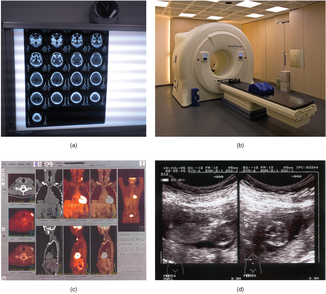
Tomography refers to imaging by sections. Computed tomography (CT) is a noninvasive imaging technique that uses computers to analyze several cross-sectional X-rays in order to reveal minute details about structures in the body ( [link] a ). The technique was invented in the 1970s and is based on the principle that, as X-rays pass through the body, they are absorbed or reflected at different levels. In the technique, a patient lies on a motorized platform while a computerized axial tomography (CAT) scanner rotates 360 degrees around the patient, taking X-ray images. A computer combines these images into a two-dimensional view of the scanned area, or “slice.”
Medical imaging techniques

These photos shows four types of imaging equipment. Photo A, the results of a CT scan, shows 17 different transverse views of the skull, each taken at a different depth along the superior-inferior axis. The images are translucent, similar to an X ray, and are viewed on a light board. Photo B shows an MRI machine, which is a large drum into which lying patients enter via a conveyor belt. Photo C shows computer images of the body taken with PET scans. This produces anterior, lateral, posterior, and transverse views of the body that reveal the structure of the internal organs. Photo D shows an ultrasound readout, which is black and white. The image depicts solid tissues as light areas and empty space as dark areas. Some of the features of a young fetus can be seen in the empty space at the center of the image. The space containing the fetus is surrounded by the solid tissue of the uterus.
(a) The results of a CT scan of the head are shown as successive transverse sections. (b) An MRI machine generates a magnetic field around a patient. (c) PET scans use radiopharmaceuticals to create images of active blood flow and physiologic activity of the organ or organs being targeted. (d) Ultrasound technology is used to monitor pregnancies because it is the least invasive of imaging techniques and uses no electromagnetic radiation. (credit a: Akira Ohgaki/flickr; credit b: “Digital Cate”/flickr; credit c: “Raziel”/Wikimedia Commons; credit d: “Isis”/Wikimedia Commons)

Since 1970, the development of more powerful computers and more sophisticated software has made CT scanning routine for many types of diagnostic evaluations. It is especially useful for soft tissue scanning, such as of the brain and the thoracic and abdominal viscera. Its level of detail is so precise that it can allow physicians to measure the size of a mass down to a millimeter. The main disadvantage of CT scanning is that it exposes patients to a dose of radiation many times higher than that of X-rays. In fact, children who undergo CT scans are at increased risk of developing cancer, as are adults who have multiple CT scans.

A CT or CAT scan relies on a circling scanner that revolves around the patient’s body. Watch this video to learn more about CT and CAT scans. What type of radiation does a CT scanner use?

Magnetic resonance imaging

Magnetic resonance imaging (MRI) is a noninvasive medical imaging technique based on a phenomenon of nuclear physics discovered in the 1930s, in which matter exposed to magnetic fields and radio waves was found to emit radio signals. In 1970, a physician and researcher named Raymond Damadian noticed that malignant (cancerous) tissue gave off different signals than normal body tissue. He applied for a patent for the first MRI scanning device, which was in use clinically by the early 1980s. The early MRI scanners were crude, but advances in digital computing and electronics led to their advancement over any other technique for precise imaging, especially to discover tumors. MRI also has the major advantage of not exposing patients to radiation.

Questions & Answers

Ayele, K., 2003. Introductory Economics, 3rd ed., Addis Ababa.
Widad Reply
can you send the book attached ?
Ariel
?
Ariel
What is economics
Widad Reply
the study of how humans make choices under conditions of scarcity
AI-Robot
U(x,y) = (x×y)1/2 find mu of x for y
Desalegn Reply
U(x,y) = (x×y)1/2 find mu of x for y
Desalegn
what is ecnomics
Jan Reply
this is the study of how the society manages it's scarce resources
Belonwu
what is macroeconomic
John Reply
macroeconomic is the branch of economics which studies actions, scale, activities and behaviour of the aggregate economy as a whole.
husaini
etc
husaini
difference between firm and industry
husaini Reply
what's the difference between a firm and an industry
Abdul
firm is the unit which transform inputs to output where as industry contain combination of firms with similar production 😅😅
Abdulraufu
Suppose the demand function that a firm faces shifted from Qd  120 3P to Qd  90  3P and the supply function has shifted from QS  20  2P to QS 10  2P . a) Find the effect of this change on price and quantity. b) Which of the changes in demand and supply is higher?
Toofiq Reply
explain standard reason why economic is a science
innocent Reply
factors influencing supply
Petrus Reply
what is economic.
Milan Reply
scares means__________________ends resources. unlimited
Jan
economics is a science that studies human behaviour as a relationship b/w ends and scares means which have alternative uses
Jan
calculate the profit maximizing for demand and supply
Zarshad Reply
Why qualify 28 supplies
Milan
what are explicit costs
Nomsa Reply
out-of-pocket costs for a firm, for example, payments for wages and salaries, rent, or materials
AI-Robot
concepts of supply in microeconomics
David Reply
economic overview notes
Amahle Reply
identify a demand and a supply curve
Salome Reply
i don't know
Parul
there's a difference
Aryan
Demand curve shows that how supply and others conditions affect on demand of a particular thing and what percent demand increase whith increase of supply of goods
Israr
Hi Sir please how do u calculate Cross elastic demand and income elastic demand?
Abari
Got questions? Join the online conversation and get instant answers!
Jobilize.com Reply

Get Jobilize Job Search Mobile App in your pocket Now!

Get it on Google Play Download on the App Store Now


Source:  OpenStax, Anatomy & Physiology. OpenStax CNX. Feb 04, 2016 Download for free at http://legacy.cnx.org/content/col11496/1.8
Google Play and the Google Play logo are trademarks of Google Inc.

Notification Switch

Would you like to follow the 'Anatomy & Physiology' conversation and receive update notifications?

Ask