<< Chapter < Page Chapter >> Page >

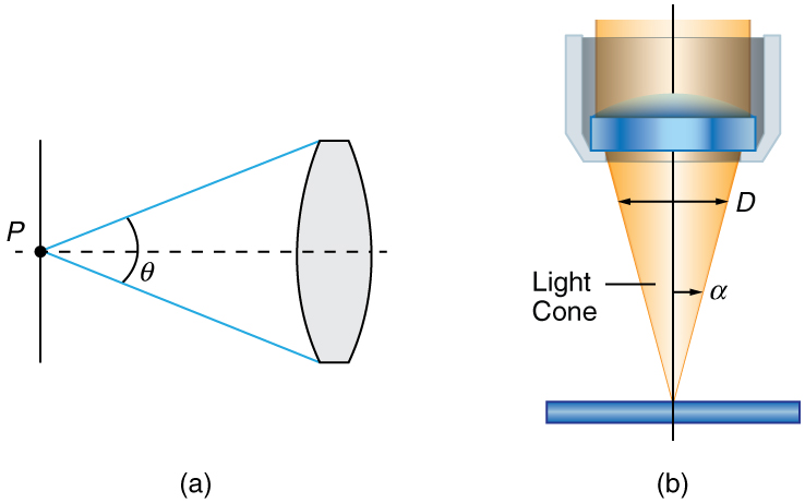
Normal optical microscopes can magnify up to 1500× with a theoretical resolution of 0.2 μm . The lenses can be quite complicated and are composed of multiple elements to reduce aberrations. Microscope objective lenses are particularly important as they primarily gather light from the specimen. Three parameters describe microscope objectives: the numerical aperture (NA) , the magnification ( m ) size 12{ \( m \) } {} , and the working distance. The NA is related to the light gathering ability of a lens and is obtained using the angle of acceptance θ size 12{θ} {} formed by the maximum cone of rays focusing on the specimen (see [link] (a)) and is given by

NA = n sin α, size 12{ ital "NA"=n"sin"α} {}

where n size 12{n} {} is the refractive index of the medium between the lens and the specimen and α = θ / 2 size 12{α= {θ} slash {2} } {} . As the angle of acceptance given by θ size 12{θ} {} increases, NA becomes larger and more light is gathered from a smaller focal region giving higher resolution. A 0 . 75 NA size 12{0 "." "75" ital "NA"} {} objective gives more detail than a 0 . 10 NA size 12{0 "." "10" ital "NA"} {} objective.

Part a of the figure shows a horizontal dotted line, a point P on the line and an objective lens at a distance from the point such that a triangle is formed from point P to the edges of the lens. An angle theta is shown at point P, representing the maximum cone of rays entering the lens from point P. Part b of the figure shows light rays from a specimen entering a camera lens held above it. The rays form an inverted cone.
(a) The numerical aperture (NA) of a microscope objective lens refers to the light-gathering ability of the lens and is calculated using half the angle of acceptance θ . (b) Here, α is half the acceptance angle for light rays from a specimen entering a camera lens, and D is the diameter of the aperture that controls the light entering the lens.

While the numerical aperture can be used to compare resolutions of various objectives, it does not indicate how far the lens could be from the specimen. This is specified by the “working distance,” which is the distance (in mm usually) from the front lens element of the objective to the specimen, or cover glass. The higher the NA size 12{ ital "NA"} {} the closer the lens will be to the specimen and the more chances there are of breaking the cover slip and damaging both the specimen and the lens. The focal length of an objective lens is different than the working distance. This is because objective lenses are made of a combination of lenses and the focal length is measured from inside the barrel. The working distance is a parameter that microscopists can use more readily as it is measured from the outermost lens. The working distance decreases as the NA size 12{ ital "NA"} {} and magnification both increase.

The term f / # size 12{ {f} slash {#} } {} in general is called the f size 12{f} {} -number and is used to denote the light per unit area reaching the image plane. In photography, an image of an object at infinity is formed at the focal point and the f size 12{f} {} -number is given by the ratio of the focal length f size 12{f} {} of the lens and the diameter D size 12{D} {} of the aperture controlling the light into the lens (see [link] (b)). If the acceptance angle is small the NA size 12{ ital "NA"} {} of the lens can also be used as given below.

f /# = f D 1 2 NA . size 12{f"/#"= { {f} over {D} } approx { {1} over {2 ital "NA"} } } {}

As the f size 12{f} {} -number decreases, the camera is able to gather light from a larger angle, giving wide-angle photography. As usual there is a trade-off. A greater f / # size 12{ {f} slash {#} } {} means less light reaches the image plane. A setting of f / 16 size 12{ {f} slash {"16"} } {} usually allows one to take pictures in bright sunlight as the aperture diameter is small. In optical fibers, light needs to be focused into the fiber. [link] shows the angle used in calculating the NA size 12{ ital "NA"} {} of an optical fiber.

Questions & Answers

what is phylogeny
Odigie Reply
evolutionary history and relationship of an organism or group of organisms
AI-Robot
ok
Deng
what is biology
Hajah Reply
the study of living organisms and their interactions with one another and their environments
AI-Robot
what is biology
Victoria Reply
HOW CAN MAN ORGAN FUNCTION
Alfred Reply
the diagram of the digestive system
Assiatu Reply
allimentary cannel
Ogenrwot
How does twins formed
William Reply
They formed in two ways first when one sperm and one egg are splited by mitosis or two sperm and two eggs join together
Oluwatobi
what is genetics
Josephine Reply
Genetics is the study of heredity
Misack
how does twins formed?
Misack
What is manual
Hassan Reply
discuss biological phenomenon and provide pieces of evidence to show that it was responsible for the formation of eukaryotic organelles
Joseph Reply
what is biology
Yousuf Reply
the study of living organisms and their interactions with one another and their environment.
Wine
discuss the biological phenomenon and provide pieces of evidence to show that it was responsible for the formation of eukaryotic organelles in an essay form
Joseph Reply
what is the blood cells
Shaker Reply
list any five characteristics of the blood cells
Shaker
lack electricity and its more savely than electronic microscope because its naturally by using of light
Abdullahi Reply
advantage of electronic microscope is easily and clearly while disadvantage is dangerous because its electronic. advantage of light microscope is savely and naturally by sun while disadvantage is not easily,means its not sharp and not clear
Abdullahi
cell theory state that every organisms composed of one or more cell,cell is the basic unit of life
Abdullahi
is like gone fail us
DENG
cells is the basic structure and functions of all living things
Ramadan
What is classification
ISCONT Reply
is organisms that are similar into groups called tara
Yamosa
in what situation (s) would be the use of a scanning electron microscope be ideal and why?
Kenna Reply
A scanning electron microscope (SEM) is ideal for situations requiring high-resolution imaging of surfaces. It is commonly used in materials science, biology, and geology to examine the topography and composition of samples at a nanoscale level. SEM is particularly useful for studying fine details,
Hilary
Got questions? Join the online conversation and get instant answers!
Jobilize.com Reply
Practice Key Terms 4

Get Jobilize Job Search Mobile App in your pocket Now!

Get it on Google Play Download on the App Store Now


Source:  OpenStax, Physics for the modern world. OpenStax CNX. Sep 16, 2015 Download for free at http://legacy.cnx.org/content/col11865/1.3
Google Play and the Google Play logo are trademarks of Google Inc.

Notification Switch

Would you like to follow the 'Physics for the modern world' conversation and receive update notifications?

Ask