<< Chapter < Page Chapter >> Page >
By the end of this section, you will be able to:
  • Identify the classes of cells that make up nervous tissue
  • Discuss how nervous tissue mediates perception and response

Nervous tissue is characterized as being excitable and capable of sending and receiving electrochemical signals that provide the body with information. Two main classes of cells make up nervous tissue: the neuron    and neuroglia    ( [link] ). Neurons send information via electrochemical impulses, called action potentials Neuroglia play an essential role in supporting neurons so they can do their very important jobs.

The neuron

This figure shows a diagram of a neuron and a micrograph showing two neuron cells. The body of the neuron contains a single, purple nucleus. The cell is irregularly shaped, having many projections emerging from its surface. Six sets of dendrites project from the top, right, and bottom edges of the cell. The dendrites are yellow and branch many times after leaving the cell, taking on the appearance of tiny trees. The axon projects from the left edge of the cell. The axon is a long cable like structure that branches into several finger like projections at its end. This is where the neuron makes contact with other cells. A label also notes that the area where the axon emerges from the cell body contains microfibrils and microtubules. The micrograph is considerably less magnified than the diagram. The neurons stain darkly and their nuclei are clearly visible. Their irregular cell body is also visible, along with the beginning of the axons.
The cell body of a neuron, also called the soma, contains the nucleus and mitochondria. The dendrites transfer the nerve impulse to the soma. The axon carries the action potential away to another excitable cell. LM × 1600. (Micrograph provided by the Regents of University of Michigan Medical School © 2012)

Follow this link to learn more about nervous tissue. What are the main parts of a nerve cell?

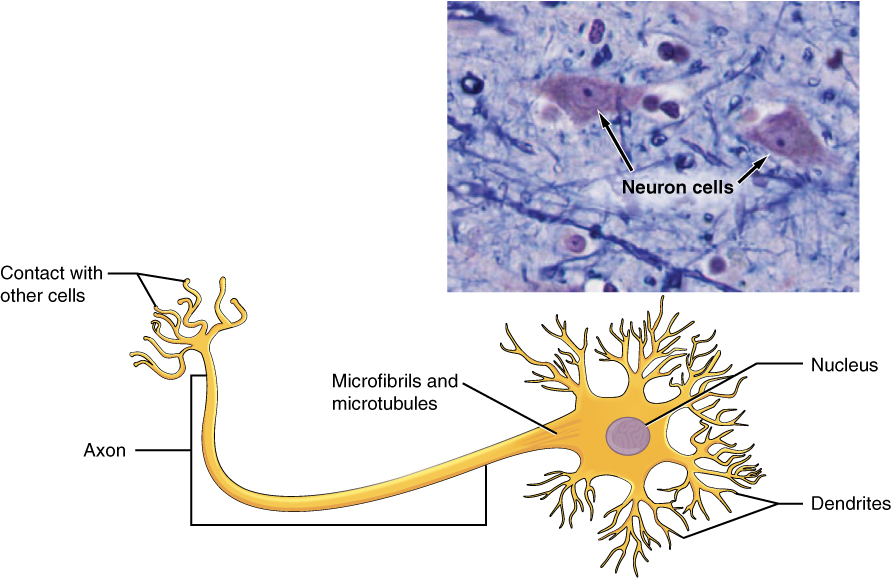
Neurons display distinctive structure, well suited to their role as conducting cells, with three main parts. The cell body includes most of the cytoplasm, the organelles, and the nucleus. Dendrites branch off the cell body and appear as thin extensions. A long “tail,” the axon, extends from the neuron body and can be wrapped in an insulating layer known as myelin    . The dendrites receive signals from other nerve cells, while the axons send the signal on to the next neuron or target organ.

The second class of neural cells comprises the neuroglia or glial cells, which have been characterized as having a simple support role. The word “glia” comes from the Greek word for glue.

Nervous tissue

Part A of this diagram shows various types of nerve cells. The largest cell is a neuron. The central body of the neuron contains a single nucleus. Six sets of dendrites project from the top, left and right, edges of the neuron. The dendrites are yellow and branch many times after leaving the cell, taking on the appearance of tiny trees. The axon projects from the bottom edge of the cell and is covered with purple sheaths labeled the myelin sheath. The sheath is not continuous, but instead is a series of equally spaced segments along the axon. Another cell, called an oligodendrocyte, is spider like in appearance, with its leg-like projections each connecting to a segment of the neuron’s myelin sheath. Above the neuron are three astrocytes. They are much smaller than the neuron and have no axons, and are also irregularly shaped cells with many dendrites projecting from the central body. Finally, a microglial cell is shown above the neuron. It is the smallest of the cells in this figure and is an elongated cell with many fine, tentacle-like projections. The projections are concentrated at the two ends of the cell, with the middle area lacking any projections. The micrograph of the neural tissue shows that this tissue is very heterogenous, with both large and small cells embedded in the matrix. Much of the space between the cells is occupied by threadlike nerve fibers.
Nervous tissue is made up of neurons and neuroglia. The cells of nervous tissue are specialized to transmit and receive impulses. LM × 872. (Micrograph provided by the Regents of University of Michigan Medical School © 2012)

Chapter review

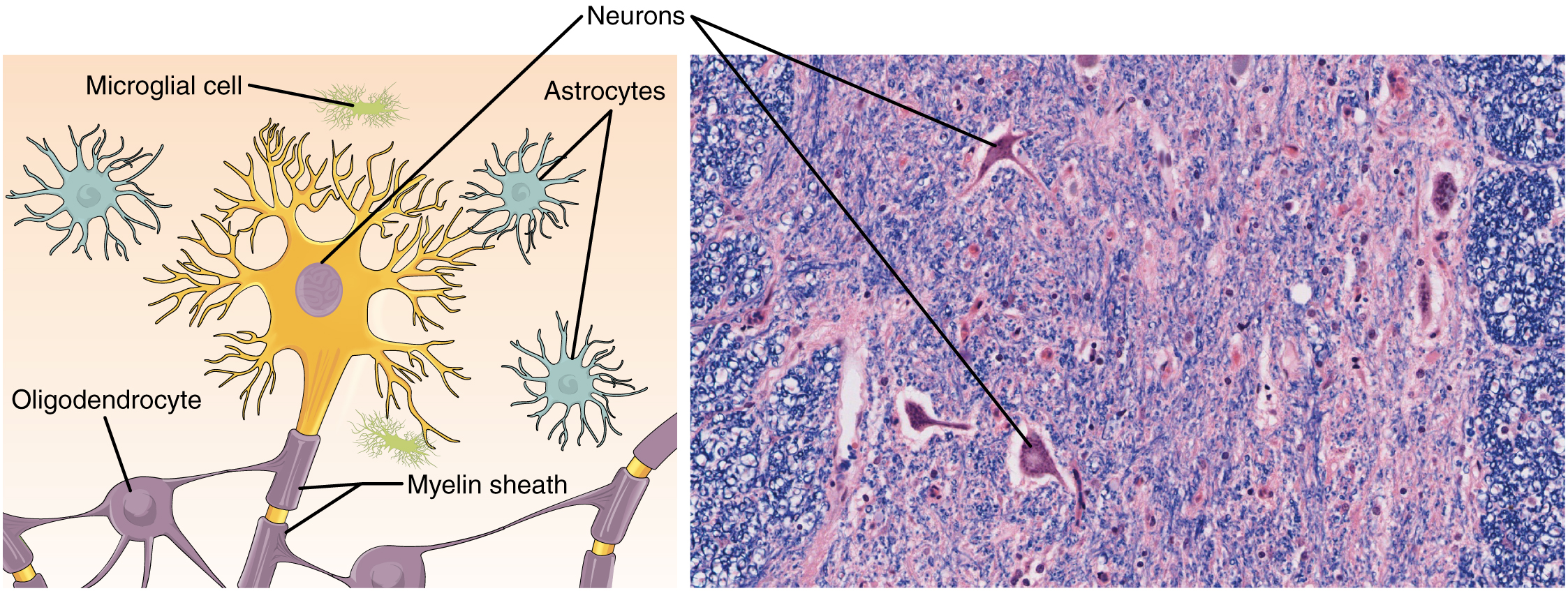
The most prominent cell of the nervous tissue, the neuron, is characterized mainly by its ability to receive stimuli and respond by generating an electrical signal, known as an action potential, which can travel rapidly over great distances in the body. A typical neuron displays a distinctive morphology: a large cell body branches out into short extensions called dendrites, which receive chemical signals from other neurons, and a long tail called an axon, which relays signals away from the cell to other neurons, muscles, or glands. Many axons are wrapped by a myelin sheath, a lipid derivative that acts as an insulator and speeds up the transmission of the action potential. Other cells in the nervous tissue, the neuroglia, include the astrocytes, microglia, oligodendrocytes, and Schwann cells.

References

Stern, P. Focus issue: getting excited about glia. Science [Internet]. 2010 [cited 2012 Dec 4]; 3(147):330-773. Available from:

(External Link)

Ming GL, Song H. Adult neurogenesis in the mammalian central nervous system. Annu. Rev. Neurosci. 2005 [cited 2012 Dec 4]; 28:223–250.

Practice Key Terms 6

Get Jobilize Job Search Mobile App in your pocket Now!

Get it on Google Play Download on the App Store Now


Source:  OpenStax, Histology. OpenStax CNX. Feb 27, 2015 Download for free at http://legacy.cnx.org/content/col11764/1.1
Google Play and the Google Play logo are trademarks of Google Inc.

Notification Switch

Would you like to follow the 'Histology' conversation and receive update notifications?

Ask