<< Chapter < Page Chapter >> Page >
Diagram of the nervous system. The central nervous system is made of the brain and spinal cord. The peripheral nervous system is made of ganglions (near the spinal cord) and nerves that run throughout the body.
The essential components of the human nervous system are shown in this illustration. The central nervous system (CNS) consists of the brain and spinal cord. It connects to the peripheral nervous system (PNS), a network of nerves that extends throughout the body.
Diagram of layers around the brain. The pia mater is a thin covering that is on the surface of the brain. Around that is cerebrospinal fluid (CSF), a region that contains blood vessels. The arachnoid maintains this space. The dura mater is the next layer out and is thick. These three layers (dura mater, arachnoid, and pia mater) make up the meninges. The next layer out is bone. The next layer is a thn periosteum, then a thin aponeurosis, and finally skin.
The layers of tissue surrounding the human brain include three meningeal membranes: the dura mater, arachnoid mater, and pia mater. (credit: modification of work by National Institutes of Health)

The blood-brain barrier

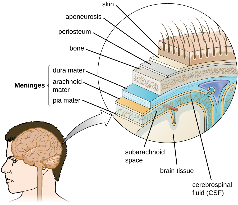
The tissues of the CNS have extra protection in that they are not exposed to blood or the immune system in the same way as other tissues. The blood vessels that supply the brain with nutrients and other chemical substances lie on top of the pia mater. The capillaries associated with these blood vessels in the brain are less permeable than those in other locations in the body. The capillary endothelial cells form tight junctions that control the transfer of blood components to the brain. In addition, cranial capillaries have far fewer fenestra (pore-like structures that are sealed by a membrane) and pinocytotic vesicles than other capillaries. As a result, materials in the circulatory system have a very limited ability to interact with the CNS directly. This phenomenon is referred to as the blood-brain barrier .

The blood-brain barrier protects the cerebrospinal fluid from contamination, and can be quite effective at excluding potential microbial pathogens. As a consequence of these defenses, there is no normal microbiota in the cerebrospinal fluid. The blood-brain barrier also inhibits the movement of many drugs into the brain, particularly compounds that are not lipid soluble. This has profound ramifications for treatments involving infections of the CNS, because it is difficult for drugs to cross the blood-brain barrier to interact with pathogens that cause infections.

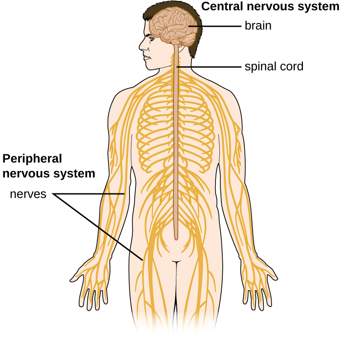
The spinal cord also has protective structures similar to those surrounding the brain. Within the bones of the vertebrae are meninges of dura mater (sometimes called the dural sheath ), arachnoid mater, pia mater, and a blood-spinal cord barrier that controls the transfer of blood components from blood vessels associated with the spinal cord.

To cause an infection in the CNS, pathogens must successfully breach the blood-brain barrier or blood-spinal cord barrier. Various pathogens employ different virulence factors and mechanisms to achieve this, but they can generally be grouped into four categories: intercellular (also called paracellular), transcellular, leukocyte facilitated, and nonhematogenous. Intercellular entry involves the use of microbial virulence factors, toxins, or inflammation-mediated processes to pass between the cells of the blood-brain barrier. In transcellular entry, the pathogen passes through the cells of the blood-brain barrier using virulence factors that allow it to adhere to and trigger uptake by vacuole- or receptor-mediated mechanisms. Leukocyte-facilitated entry is a Trojan-horse mechanism that occurs when a pathogen infects peripheral blood leukocytes to directly enter the CNS. Nonhematogenous entry allows pathogens to enter the brain without encountering the blood-brain barrier; it occurs when pathogens travel along either the olfactory or trigeminal cranial nerves that lead directly into the CNS.

Questions & Answers

what is biology
Hajah Reply
the study of living organisms and their interactions with one another and their environments
AI-Robot
what is biology
Victoria Reply
HOW CAN MAN ORGAN FUNCTION
Alfred Reply
the diagram of the digestive system
Assiatu Reply
allimentary cannel
Ogenrwot
How does twins formed
William Reply
They formed in two ways first when one sperm and one egg are splited by mitosis or two sperm and two eggs join together
Oluwatobi
what is genetics
Josephine Reply
Genetics is the study of heredity
Misack
how does twins formed?
Misack
What is manual
Hassan Reply
discuss biological phenomenon and provide pieces of evidence to show that it was responsible for the formation of eukaryotic organelles
Joseph Reply
what is biology
Yousuf Reply
the study of living organisms and their interactions with one another and their environment.
Wine
discuss the biological phenomenon and provide pieces of evidence to show that it was responsible for the formation of eukaryotic organelles in an essay form
Joseph Reply
what is the blood cells
Shaker Reply
list any five characteristics of the blood cells
Shaker
lack electricity and its more savely than electronic microscope because its naturally by using of light
Abdullahi Reply
advantage of electronic microscope is easily and clearly while disadvantage is dangerous because its electronic. advantage of light microscope is savely and naturally by sun while disadvantage is not easily,means its not sharp and not clear
Abdullahi
cell theory state that every organisms composed of one or more cell,cell is the basic unit of life
Abdullahi
is like gone fail us
DENG
cells is the basic structure and functions of all living things
Ramadan
What is classification
ISCONT Reply
is organisms that are similar into groups called tara
Yamosa
in what situation (s) would be the use of a scanning electron microscope be ideal and why?
Kenna Reply
A scanning electron microscope (SEM) is ideal for situations requiring high-resolution imaging of surfaces. It is commonly used in materials science, biology, and geology to examine the topography and composition of samples at a nanoscale level. SEM is particularly useful for studying fine details,
Hilary
cell is the building block of life.
Condoleezza Reply
Got questions? Join the online conversation and get instant answers!
Jobilize.com Reply

Get Jobilize Job Search Mobile App in your pocket Now!

Get it on Google Play Download on the App Store Now


Source:  OpenStax, Microbiology. OpenStax CNX. Nov 01, 2016 Download for free at http://cnx.org/content/col12087/1.4
Google Play and the Google Play logo are trademarks of Google Inc.

Notification Switch

Would you like to follow the 'Microbiology' conversation and receive update notifications?

Ask