

| << Chapter < Page | Chapter >> Page > |
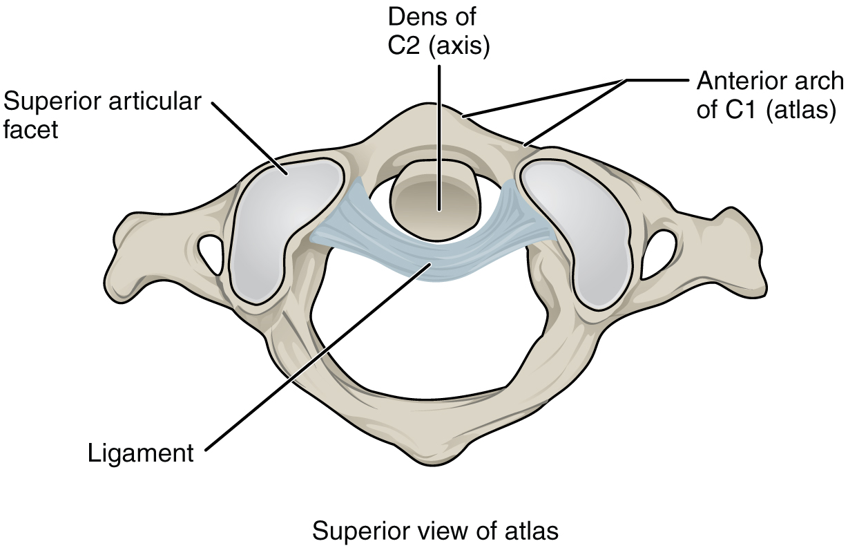
The atlantoaxial joint , between the atlas and axis, consists of three articulations. The paired superior articular processes of the axis articulate with the inferior articular processes of the atlas. These articulating surfaces are relatively flat and oriented horizontally. The third articulation is the pivot joint formed between the dens, which projects upward from the body of the axis, and the inner aspect of the anterior arch of the atlas ( [link] ). A strong ligament passes posterior to the dens to hold it in position against the anterior arch. These articulations allow the atlas to rotate on top of the axis, moving the head toward the right or left, as when shaking your head “no.”
The temporomandibular joint (TMJ) is the joint that allows for opening (mandibular depression) and closing (mandibular elevation) of the mouth, as well as side-to-side and protraction/retraction motions of the lower jaw. This joint involves the articulation between the mandibular fossa and articular tubercle of the temporal bone, with the condyle (head) of the mandible. Located between these bony structures, filling the gap between the skull and mandible, is a flexible articular disc ( [link] ). This disc serves to smooth the movements between the temporal bone and mandibular condyle.
Movement at the TMJ during opening and closing of the mouth involves both gliding and hinge motions of the mandible. With the mouth closed, the mandibular condyle and articular disc are located within the mandibular fossa of the temporal bone. During opening of the mouth, the mandible hinges downward and at the same time is pulled anteriorly, causing both the condyle and the articular disc to glide forward from the mandibular fossa onto the downward projecting articular tubercle. The net result is a forward and downward motion of the condyle and mandibular depression. The temporomandibular joint is supported by an extrinsic ligament that anchors the mandible to the skull. This ligament spans the distance between the base of the skull and the lingula on the medial side of the mandibular ramus.
Dislocation of the TMJ may occur when opening the mouth too wide (such as when taking a large bite) or following a blow to the jaw, resulting in the mandibular condyle moving beyond (anterior to) the articular tubercle. In this case, the individual would not be able to close his or her mouth. Temporomandibular joint disorder is a painful condition that may arise due to arthritis, wearing of the articular cartilage covering the bony surfaces of the joint, muscle fatigue from overuse or grinding of the teeth, damage to the articular disc within the joint, or jaw injury. Temporomandibular joint disorders can also cause headache, difficulty chewing, or even the inability to move the jaw (lock jaw). Pharmacologic agents for pain or other therapies, including bite guards, are used as treatments.
Notification Switch
Would you like to follow the 'Anatomy & Physiology' conversation and receive update notifications?

|
|
|
|
|

|
|

|
|

|
|

|
|

|
|

|
|

|
|

|
|

|
|

|