<< Chapter < Page Chapter >> Page >

Thigh muscles that move the femur, tibia, and fibula

Deep fascia in the thigh separates it into medial, anterior, and posterior compartments (see [link] and [link] ). The muscles in the medial compartment of the thigh    are responsible for adducting the femur at the hip. Along with the adductor longus, adductor brevis, adductor magnus, and pectineus, the strap-like gracilis    adducts the thigh in addition to flexing the leg at the knee.

Thigh muscles that move the femur, tibia, and fibula

This table describes the thigh muscles that move the femur, tibia, and fibula. The medial compartment of the thigh consists of the gracilis, which moves the back of the lower legs up toward the buttocks, as when kneeling; it also assists in opening the thighs. It originates in the inferior ramus, the body of the pubis, and the ischial ramus. These muscles, the quadriceps femoris group, make up the anterior compartment of the thigh. The rectus femoris moves the lower leg out in front of the body, as when kicking; it also assists in raising the knee. It originates in the anterior inferior iliac spine and in the superior margin of the acetabulum. The vastus lateralis moves the lower leg out in front of the body, as when kicking. It originates in the greater trochanter, the intertrochanteric line, and the linea aspera. The vastus medialis moves the lower leg out in front of the body, as when kicking. It originates in the linea aspera and the intertrochanteric line. The vastus intermedius moves the lower leg out in front of the body, as when kicking. It originates in the proximal femur shaft. The sartorius moves the back of the lower legs up and back toward the buttocks, as when kneeling; it also assists in moving the thigh diagonally upward and outward as when mounting a bike. It originates in the anterior superior iliac spine. These muscles, the hamstring group, make up the posterior compartment of the thigh. The biceps femoris moves the back of the lower leg up and back toward the buttocks, as when kneeling; it also moves the thigh down and back and twists the thigh (and lower leg) outward. It originates in the ischial tuberosity, linea aspera, and distal femur. The semitendinosus moves the back of the lower legs up toward the buttocks, as when kneeling; it also moves the thigh down and back and twists the thigh (and lower leg) inward. It originates in the ischial tuberosity. The semi-membranosus moves the back of the lower legs up and back toward the buttocks, as when kneeling; it also moves the thigh down and back and twists the thigh (and lower leg) inward. It originates in the ischial tuberosity.

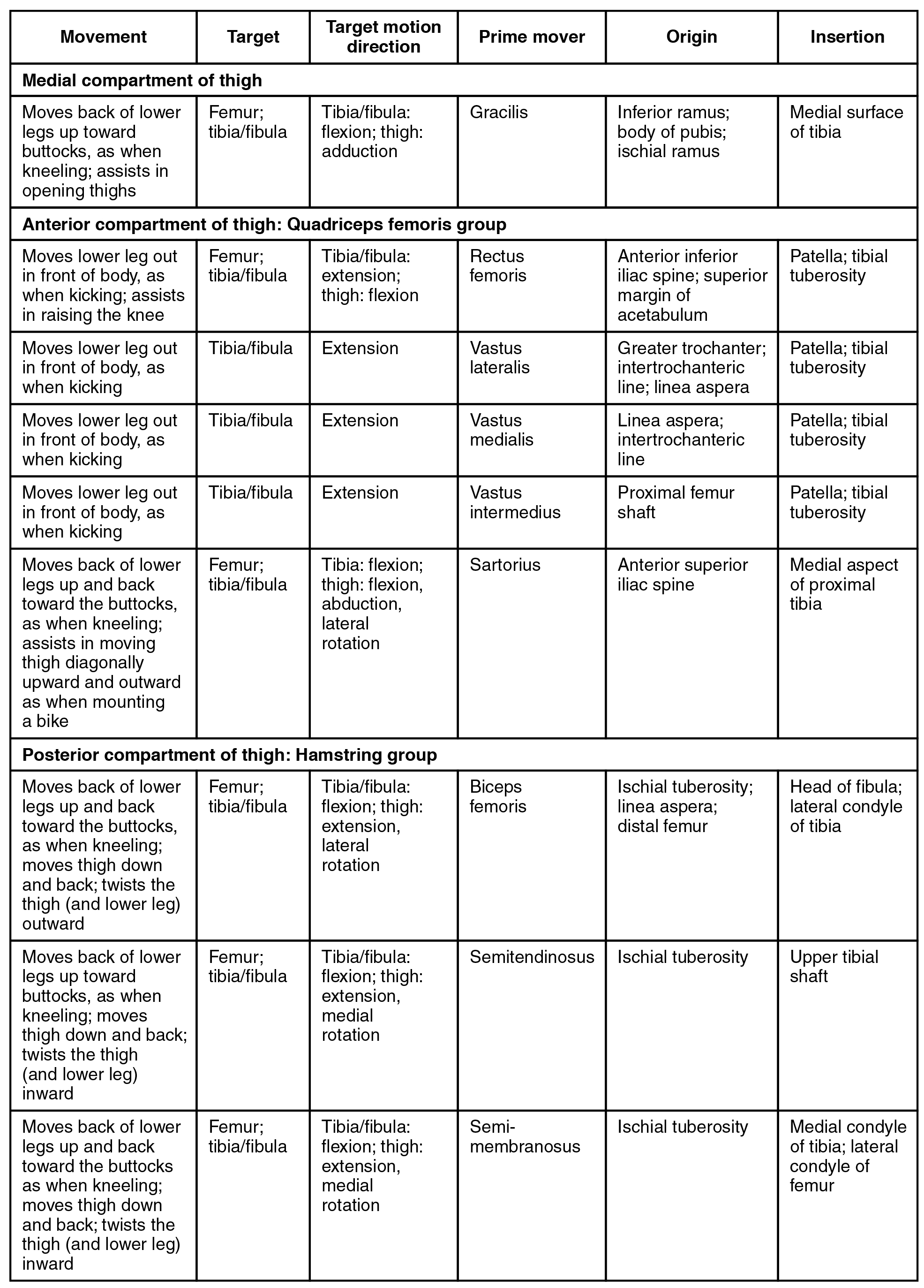
The muscles of the anterior compartment of the thigh    flex the thigh and extend the leg. This compartment contains the quadriceps femoris group    , which actually comprises four muscles that extend and stabilize the knee. The rectus femoris    is on the anterior aspect of the thigh, the vastus lateralis    is on the lateral aspect of the thigh, the vastus medialis    is on the medial aspect of the thigh, and the vastus intermedius    is between the vastus lateralis and vastus medialis and deep to the rectus femoris. The tendon common to all four is the quadriceps tendon    (patellar tendon), which inserts into the patella and continues below it as the patellar ligament    . The patellar ligament attaches to the tibial tuberosity. In addition to the quadriceps femoris, the sartorius    is a band-like muscle that extends from the anterior superior iliac spine to the medial side of the proximal tibia. This versatile muscle flexes the leg at the knee and flexes, abducts, and laterally rotates the leg at the hip. This muscle allows us to sit cross-legged.

The posterior compartment of the thigh    includes muscles that flex the leg and extend the thigh. The three long muscles on the back of the knee are the hamstring group    , which flexes the knee. These are the biceps femoris    , semitendinosus    , and semimembranosus    . The tendons of these muscles form the popliteal fossa    , the diamond-shaped space at the back of the knee.

Muscles that move the feet and toes

Similar to the thigh muscles, the muscles of the leg are divided by deep fascia into compartments, although the leg has three: anterior, lateral, and posterior ( [link] and [link] ).

Muscles of the lower leg

The left panel shows the superficial muscles that move the feet and the center panel shows the posterior view of the same muscles. The right panel shows the deep muscles of the right lower leg.
The muscles of the anterior compartment of the lower leg are generally responsible for dorsiflexion, and the muscles of the posterior compartment of the lower leg are generally responsible for plantar flexion. The lateral and medial muscles in both compartments invert, evert, and rotate the foot.

Muscles that move the feet and toes

This tables describes the muscles that move the feet and toes. These muscles make up the anterior compartment of the leg. The tibialis anterior raises the sole of the foot off the ground, as when preparing to foot-tap; it also bends the inside of the foot upwards, as when catching your balance while falling laterally toward the opposite side as the balancing foot. It originates in the lateral condyle and upper tibial shaft and in the interosseous membrane. The extensor hallucis longus raises the sole of the foot off the ground, as when preparing to foot-tap; it also extends the big toe. It originates in the anteromedial fibula shaft and interosseous membrane. The extensor digitorum longus raises the sole of the foot off the ground, as when preparing to foot-tap; it also extends the toes. It originates in the lateral condyle of the tibia, the proximal portion of the fibula, and the interosseous membrane. These muscles make up the lateral compartment of the leg. The fibularis longus lowers the sole of the foot to the ground, as when foot-tapping or jumping; it also bends the inside of the foot downwards, as when catching your balance while falling laterally toward the same side as the balancing foot. It originates in the upper portion of the lateral fibula. The fibularis (peroneus) brevis lowers the side of the foot to the ground, as when foot-tapping or jumping; it also bends the inside of the foot downward, as when catching your balance while falling laterally toward the same side as the balancing foot. It originates in the distal fibula shaft. These superficial muscles make up the posterior compartment of the leg. The gastrocnemius lowers the sole of the foot to the ground, as when foot-tapping or jumping; it also assists in moving the back of the lower legs up and back toward the buttocks. It originates in the medial and lateral condyles of the femur. The soleus lowers the sole of the foot the ground, as when foot-tapping or jumping; it also maintains posture while walking. It originates in the superior tibia, fibula, and interosseous membrane. The plantaris lowers the sole of the foot to the ground, as when foot-tapping or jumping; it also assists in moving the back of the lower legs up and back toward the buttocks. It originates in the posterior femur above the lateral condyle. The tibialis posterior lowers the sole of the foot to the ground, as when foot-tapping or jumping. It originates in the superior tibia and fibula and in the interosseous membrane. These deep muscles also make up the posterior compartment of the leg. The popliteus moves the back of the lower legs up and back toward the buttocks; it also assists in rotation of the leg at the knee and thigh. It originates in the lateral condyle of the femur and the lateral meniscus. The flexor digitorum longus lowers the sole of the foot to the ground, as when foot-tapping or jumping; it also bends the inside of the foot upward and flexes the toes. It originates in the posterior tibia. The flexor hallicis longus flexes the big toe. It originates in the midshaft of the fibula and in the interosseous membrane.

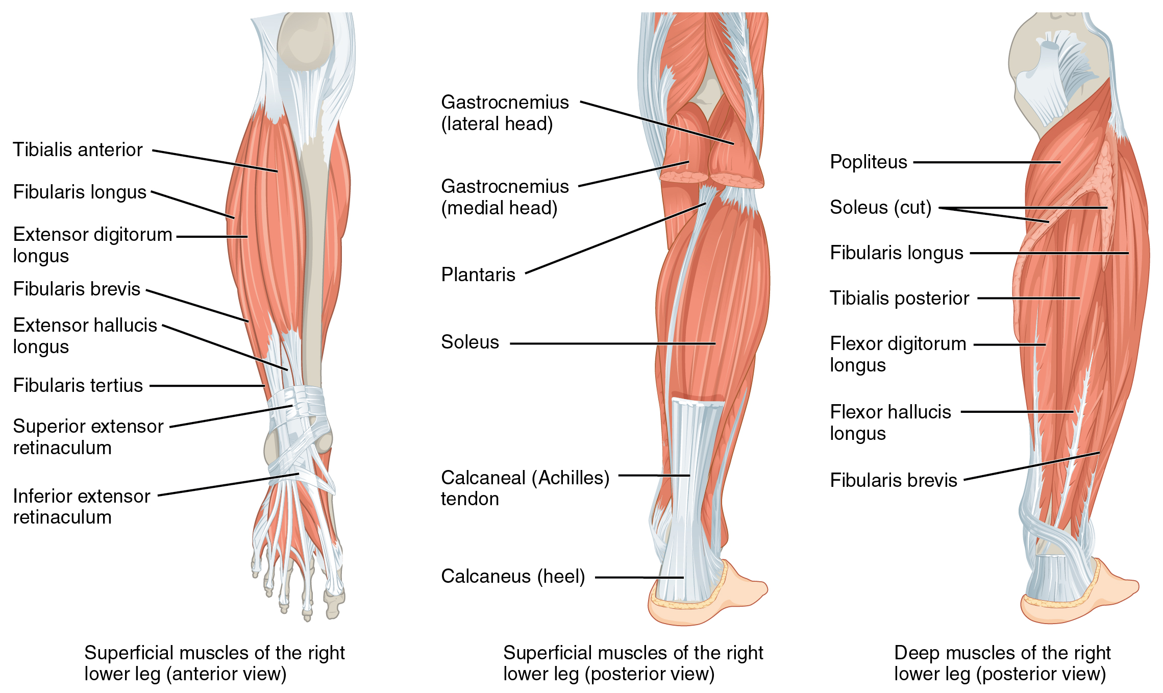
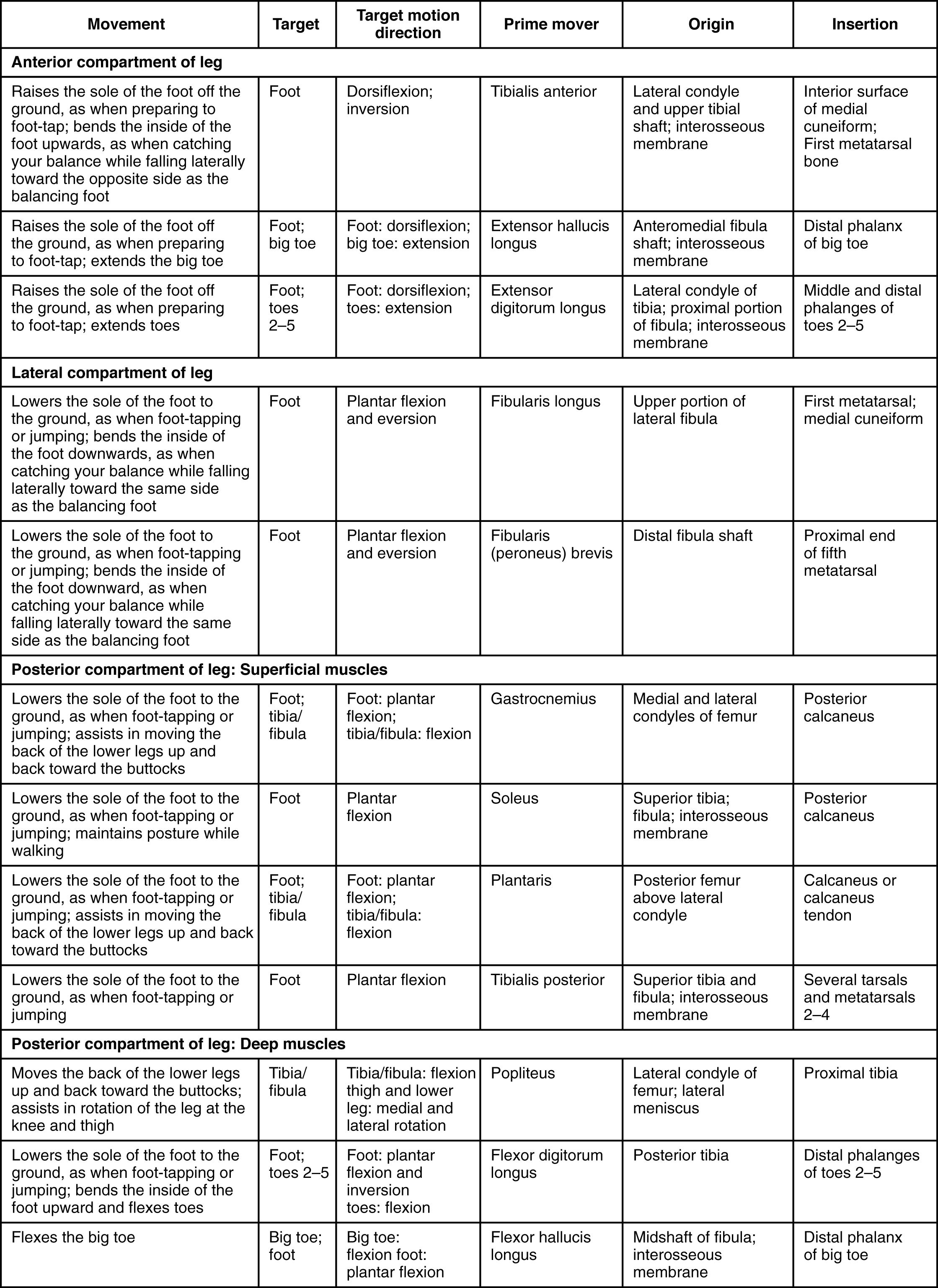
The muscles in the anterior compartment of the leg    : the tibialis anterior    , a long and thick muscle on the lateral surface of the tibia, the extensor hallucis longus    , deep under it, and the extensor digitorum longus    , lateral to it, all contribute to raising the front of the foot when they contract. The fibularis tertius    , a small muscle that originates on the anterior surface of the fibula, is associated with the extensor digitorum longus and sometimes fused to it, but is not present in all people. Thick bands of connective tissue called the superior extensor retinaculum    (transverse ligament of the ankle) and the inferior extensor retinaculum    , hold the tendons of these muscles in place during dorsiflexion.

Questions & Answers

how does Neisseria cause meningitis
Nyibol Reply
what is microbiologist
Muhammad Reply
what is errata
Muhammad
is the branch of biology that deals with the study of microorganisms.
Ntefuni Reply
What is microbiology
Mercy Reply
studies of microbes
Louisiaste
when we takee the specimen which lumbar,spin,
Ziyad Reply
How bacteria create energy to survive?
Muhamad Reply
Bacteria doesn't produce energy they are dependent upon their substrate in case of lack of nutrients they are able to make spores which helps them to sustain in harsh environments
_Adnan
But not all bacteria make spores, l mean Eukaryotic cells have Mitochondria which acts as powerhouse for them, since bacteria don't have it, what is the substitution for it?
Muhamad
they make spores
Louisiaste
what is sporadic nd endemic, epidemic
Aminu Reply
the significance of food webs for disease transmission
Abreham
food webs brings about an infection as an individual depends on number of diseased foods or carriers dully.
Mark
explain assimilatory nitrate reduction
Esinniobiwa Reply
Assimilatory nitrate reduction is a process that occurs in some microorganisms, such as bacteria and archaea, in which nitrate (NO3-) is reduced to nitrite (NO2-), and then further reduced to ammonia (NH3).
Elkana
This process is called assimilatory nitrate reduction because the nitrogen that is produced is incorporated in the cells of microorganisms where it can be used in the synthesis of amino acids and other nitrogen products
Elkana
Examples of thermophilic organisms
Shu Reply
Give Examples of thermophilic organisms
Shu
advantages of normal Flora to the host
Micheal Reply
Prevent foreign microbes to the host
Abubakar
they provide healthier benefits to their hosts
ayesha
They are friends to host only when Host immune system is strong and become enemies when the host immune system is weakened . very bad relationship!
Mark
what is cell
faisal Reply
cell is the smallest unit of life
Fauziya
cell is the smallest unit of life
Akanni
ok
Innocent
cell is the structural and functional unit of life
Hasan
is the fundamental units of Life
Musa
what are emergency diseases
Micheal Reply
There are nothing like emergency disease but there are some common medical emergency which can occur simultaneously like Bleeding,heart attack,Breathing difficulties,severe pain heart stock.Hope you will get my point .Have a nice day ❣️
_Adnan
define infection ,prevention and control
Innocent
I think infection prevention and control is the avoidance of all things we do that gives out break of infections and promotion of health practices that promote life
Lubega
Heyy Lubega hussein where are u from?
_Adnan
en français
Adama
which site have a normal flora
ESTHER Reply
Many sites of the body have it Skin Nasal cavity Oral cavity Gastro intestinal tract
Safaa
skin
Asiina
skin,Oral,Nasal,GIt
Sadik
How can Commensal can Bacteria change into pathogen?
Sadik
How can Commensal Bacteria change into pathogen?
Sadik
all
Tesfaye
by fussion
Asiina
what are the advantages of normal Flora to the host
Micheal
what are the ways of control and prevention of nosocomial infection in the hospital
Micheal
what is inflammation
Shelly Reply
part of a tissue or an organ being wounded or bruised.
Wilfred
what term is used to name and classify microorganisms?
Micheal Reply
Binomial nomenclature
adeolu
Got questions? Join the online conversation and get instant answers!
Jobilize.com Reply

Get Jobilize Job Search Mobile App in your pocket Now!

Get it on Google Play Download on the App Store Now


Source:  OpenStax, Anatomy & Physiology. OpenStax CNX. Feb 04, 2016 Download for free at http://legacy.cnx.org/content/col11496/1.8
Google Play and the Google Play logo are trademarks of Google Inc.

Notification Switch

Would you like to follow the 'Anatomy & Physiology' conversation and receive update notifications?

Ask