<< Chapter < Page Chapter >> Page >
By the end of this section, you will be able to:
  • Describe the structures found in the PNS
  • Distinguish between somatic and autonomic structures, including the special peripheral structures of the enteric nervous system
  • Name the twelve cranial nerves and explain the functions associated with each
  • Describe the sensory and motor components of spinal nerves and the plexuses that they pass through

The PNS is not as contained as the CNS because it is defined as everything that is not the CNS. Some peripheral structures are incorporated into the other organs of the body. In describing the anatomy of the PNS, it is necessary to describe the common structures, the nerves and the ganglia, as they are found in various parts of the body. Many of the neural structures that are incorporated into other organs are features of the digestive system; these structures are known as the enteric nervous system    and are a special subset of the PNS.

Ganglia

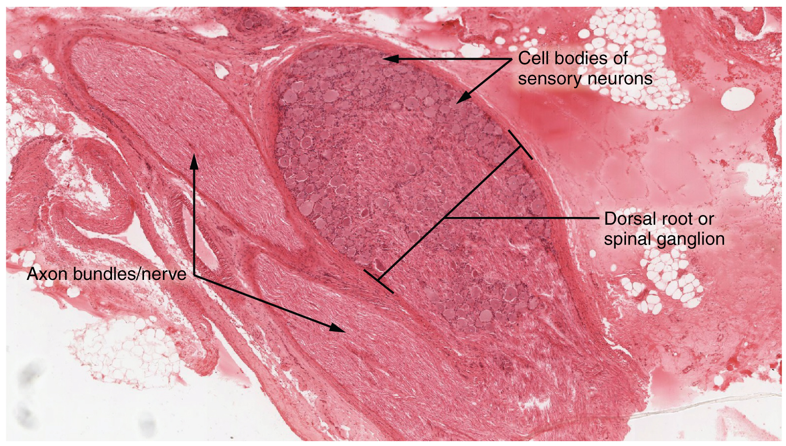
A ganglion is a group of neuron cell bodies in the periphery. Ganglia can be categorized, for the most part, as either sensory ganglia or autonomic ganglia, referring to their primary functions. The most common type of sensory ganglion is a dorsal (posterior) root ganglion    . These ganglia are the cell bodies of neurons with axons that are sensory endings in the periphery, such as in the skin, and that extend into the CNS through the dorsal nerve root. The ganglion is an enlargement of the nerve root. Under microscopic inspection, it can be seen to include the cell bodies of the neurons, as well as bundles of fibers that are the posterior nerve root ( [link] ). The cells of the dorsal root ganglion are unipolar cells, classifying them by shape. Also, the small round nuclei of satellite cells can be seen surrounding—as if they were orbiting—the neuron cell bodies.

Dorsal root ganglion

This micrograph shows the structure of the dorsal root ganglion. The cell bodies of the neurons and the axon bundles are also labeled.
The cell bodies of sensory neurons, which are unipolar neurons by shape, are seen in this photomicrograph. Also, the fibrous region is composed of the axons of these neurons that are passing through the ganglion to be part of the dorsal nerve root (tissue source: canine). LM × 40. (Micrograph provided by the Regents of University of Michigan Medical School © 2012)

Spinal cord and root ganglion

This micrograph shows a magnified view of the dorsal root ganglion, showing the satellite cells and the cell bodies of sensory neurons.
The slide includes both a cross-section of the lumbar spinal cord and a section of the dorsal root ganglion (see also [link] ) (tissue source: canine). LM × 1600. (Micrograph provided by the Regents of University of Michigan Medical School © 2012)

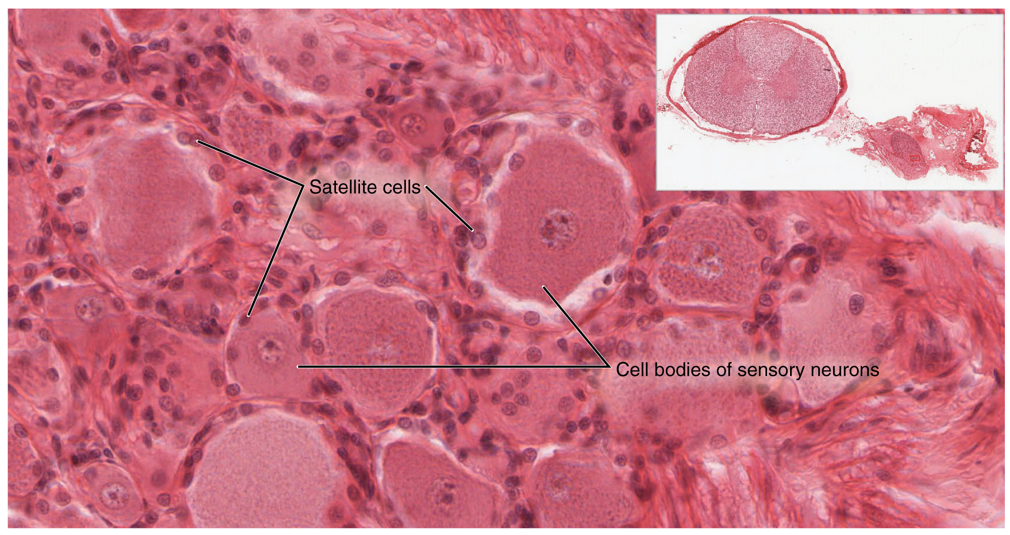
View the University of Michigan WebScope at (External Link) to explore the tissue sample in greater detail. If you zoom in on the dorsal root ganglion, you can see smaller satellite glial cells surrounding the large cell bodies of the sensory neurons. From what structure do satellite cells derive during embryologic development?

Another type of sensory ganglion is a cranial nerve ganglion    . This is analogous to the dorsal root ganglion, except that it is associated with a cranial nerve    instead of a spinal nerve    . The roots of cranial nerves are within the cranium, whereas the ganglia are outside the skull. For example, the trigeminal ganglion    is superficial to the temporal bone whereas its associated nerve is attached to the mid-pons region of the brain stem. The neurons of cranial nerve ganglia are also unipolar in shape with associated satellite cells.

Questions & Answers

what is cell
Oppicial Reply
To know how bones are functions
DAUDA Reply
diagram of the heart
Victoria Reply
what are the layers of the muscles
Tongdock Reply
What is Amebae
Najibu Reply
the collection of fluids in the throat is cause by what
Emmanuel Reply
what is difference between meiosis and mitosis
Aishetu Reply
what is difference between mitosis and meiosis
Aishetu
What is Anatomy
Najibu Reply
What the difference between the Anatomy and physiology
Najibu
What is the meaning of chromoprotein
Aisha Reply
what is cartilage
Abdulkadir Reply
tough , white fibrous tissue
Henry
distinguish between anatomy and physiology
Amina Reply
Anatomy is the study of internal structure of an organism while physiology is the study of the function/relationship of the body organs working together as a system in an organism.
adeyeye
distinguish between anatomy and physiology
Erny Reply
regional anatomy is the study of the body regionally
Ismail Reply
what is the meaning of regional anatomy
Aminat Reply
epithelial tissue: it covers the Hollow organs and body cavities
Esomchi Reply

Get Jobilize Job Search Mobile App in your pocket Now!

Get it on Google Play Download on the App Store Now


Source:  OpenStax, Anatomy & Physiology. OpenStax CNX. Feb 04, 2016 Download for free at http://legacy.cnx.org/content/col11496/1.8
Google Play and the Google Play logo are trademarks of Google Inc.

Notification Switch

Would you like to follow the 'Anatomy & Physiology' conversation and receive update notifications?

Ask