

| << Chapter < Page | Chapter >> Page > |
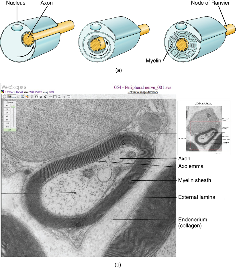
The appearance of the myelin sheath can be thought of as similar to the pastry wrapped around a hot dog for “pigs in a blanket” or a similar food. The glial cell is wrapped around the axon several times with little to no cytoplasm between the glial cell layers. For oligodendrocytes, the rest of the cell is separate from the myelin sheath as a cell process extends back toward the cell body. A few other processes provide the same insulation for other axon segments in the area. For Schwann cells, the outermost layer of the cell membrane contains cytoplasm and the nucleus of the cell as a bulge on one side of the myelin sheath. During development, the glial cell is loosely or incompletely wrapped around the axon ( [link] a ). The edges of this loose enclosure extend toward each other, and one end tucks under the other. The inner edge wraps around the axon, creating several layers, and the other edge closes around the outside so that the axon is completely enclosed.
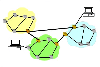
View the University of Michigan WebScope at (External Link)& to see an electron micrograph of a cross-section of a myelinated nerve fiber. The axon contains microtubules and neurofilaments that are bounded by a plasma membrane known as the axolemma. Outside the plasma membrane of the axon is the myelin sheath, which is composed of the tightly wrapped plasma membrane of a Schwann cell. What aspects of the cells in this image react with the stain to make them a deep, dark, black color, such as the multiple layers that are the myelin sheath?
Myelin sheaths can extend for one or two millimeters, depending on the diameter of the axon. Axon diameters can be as small as 1 to 20 micrometers. Because a micrometer is 1/1000 of a millimeter, this means that the length of a myelin sheath can be 100–1000 times the diameter of the axon. [link] , [link] , and [link] show the myelin sheath surrounding an axon segment, but are not to scale. If the myelin sheath were drawn to scale, the neuron would have to be immense—possibly covering an entire wall of the room in which you are sitting.
Multiple sclerosis (MS) is one such disease. It is an example of an autoimmune disease. The antibodies produced by lymphocytes (a type of white blood cell) mark myelin as something that should not be in the body. This causes inflammation and the destruction of the myelin in the central nervous system. As the insulation around the axons is destroyed by the disease, scarring becomes obvious. This is where the name of the disease comes from; sclerosis means hardening of tissue, which is what a scar is. Multiple scars are found in the white matter of the brain and spinal cord. The symptoms of MS include both somatic and autonomic deficits. Control of the musculature is compromised, as is control of organs such as the bladder.
Guillain-Barré (pronounced gee-YAN bah-RAY) syndrome is an example of a demyelinating disease of the peripheral nervous system. It is also the result of an autoimmune reaction, but the inflammation is in peripheral nerves. Sensory symptoms or motor deficits are common, and autonomic failures can lead to changes in the heart rhythm or a drop in blood pressure, especially when standing, which causes dizziness.
Nervous tissue contains two major cell types, neurons and glial cells. Neurons are the cells responsible for communication through electrical signals. Glial cells are supporting cells, maintaining the environment around the neurons.
Neurons are polarized cells, based on the flow of electrical signals along their membrane. Signals are received at the dendrites, are passed along the cell body, and propagate along the axon towards the target, which may be another neuron, muscle tissue, or a gland. Many axons are insulated by a lipid-rich substance called myelin. Specific types of glial cells provide this insulation.
Several types of glial cells are found in the nervous system, and they can be categorized by the anatomical division in which they are found. In the CNS, astrocytes, oligodendrocytes, microglia, and ependymal cells are found. Astrocytes are important for maintaining the chemical environment around the neuron and are crucial for regulating the blood-brain barrier. Oligodendrocytes are the myelinating glia in the CNS. Microglia act as phagocytes and play a role in immune surveillance. Ependymal cells are responsible for filtering the blood to produce cerebrospinal fluid, which is a circulatory fluid that performs some of the functions of blood in the brain and spinal cord because of the BBB. In the PNS, satellite cells are supporting cells for the neurons, and Schwann cells insulate peripheral axons.
Visit this site to learn about how nervous tissue is composed of neurons and glial cells. The neurons are dynamic cells with the ability to make a vast number of connections and to respond incredibly quickly to stimuli and to initiate movements based on those stimuli. They are the focus of intense research as failures in physiology can lead to devastating illnesses. Why are neurons only found in animals? Based on what this article says about neuron function, why wouldn’t they be helpful for plants or microorganisms?
Neurons enable thought, perception, and movement. Plants do not move, so they do not need this type of tissue. Microorganisms are too small to have a nervous system. Many are single-celled, and therefore have organelles for perception and movement.
View the University of Michigan WebScope at (External Link)& to see an electron micrograph of a cross-section of a myelinated nerve fiber. The axon contains microtubules and neurofilaments, bounded by a plasma membrane known as the axolemma. Outside the plasma membrane of the axon is the myelin sheath, which is composed of the tightly wrapped plasma membrane of a Schwann cell. What aspects of the cells in this image react with the stain that makes them the deep, dark, black color, such as the multiple layers that are the myelin sheath?
Lipid membranes, such as the cell membrane and organelle membranes.
Notification Switch
Would you like to follow the 'Anatomy & Physiology' conversation and receive update notifications?

|
|
|
|
|
|
|

|
|

|
|

|
|

|
|

|
|

|
|

|
|

|
|

|