Determination of molecular weight of polymers and proteins in the range of 3.5 kDa to 2 MDa by correlating molecular weight and mobility diameter.
Determination of absolute number concentration of nanoparticles in solution by obtaining the ES droplet size distributions and using statistical analysis to find the original monomer concentration. Dimers or trimers can be formed in the electrospray process due to droplet induced aggregation and are observed in the spectrum.
Kinetics of aggregation of nanoparticles in solution by analysis of multimodal mobility distributions from which distinct types of aggregation states can be identified.
Quantification of ligand adsorption to bionanoparticles by measuring the reduction in electrical mobility of a complex particle (particle-protein) that corresponds to an increase in mobility diameter.
Characterization of sam-functionalized gold nanoparticles by es-dma
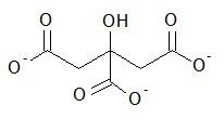
Citrate (
[link] ) stabilized gold nanoparticles (AuNPs) with diameter in the range 10-60 nm and conjugated AuNPs are analyzed by ES-DMA. This investigation shows that the formation of salt particles on the surface of AuNPs can interfere with the mobility analysis because of the reduction in analyte signals. Since sodium citrate is a non volatile soluble salt, ES produces two types of droplets. One droplet consists of AuNPs and salt and the other droplet contains only salt. Thus, samples must be cleaned by centrifugation prior to determine the size of bare AuNPs.
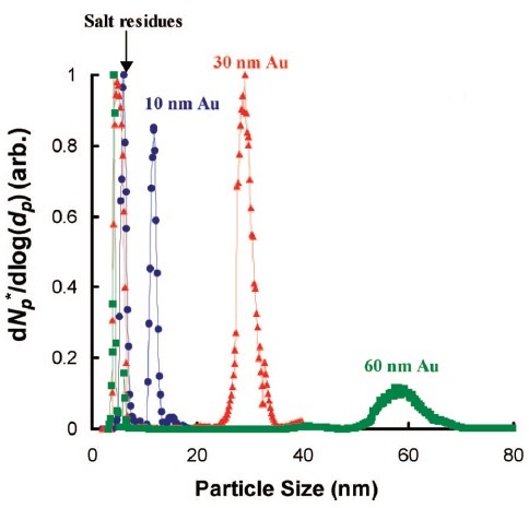
[link] presents the size distribution of AuNPs of distinct diameters and peaks corresponding to salt residues.
Structure of citrate that provides charge stabilization to AuNPs.Particle size distribution of 10 nm, 30 nm and 60 nm AuNPs after centrifugation cleaning. Reprinted with permission from D. Tsai, R. A. Zangmeister, L. F. Pease III, M. J. Tarlov and M. R. Zachariah.
Langmuir , 2008,
24 , 8483. Copyright (2015) American Chemical Society.
The mobility size of bare AuNPs (d
p0 ) can be obtained by using
[link] , where d
p,m and d
s are mobility sizes of the AuNPs encrusted with salts and the salt NP, respectively. However, the presence of self-assembled monolayer (SAM) produces a difference in electrical mobility between conjugated and bare AuNPs. Hence, the determination of the diameter of AuNPs (salt-free) is critical to distinguish the increment in size after functionalization with SAM. The coating thickness of SAM that corresponds to the change in particle size (ΔL) is calculated by using
[link] , where d
p and d
p0 are the coated and uncoated particle mobility diameters, respectively.
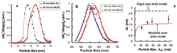
In addition, the change in particle size can be determined by considering a simple rigid core-shell model that gives theoretical values of ΔL
1 higher than the experimental ones (ΔL). A modified core-shell model is proposed in which a size dependent effect on ΔL
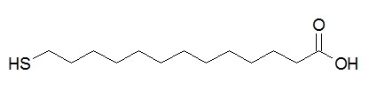
2 is observed for a range of particle sizes. AuNPs of 10 nm and 60 nm are coated with MUA (
[link] ), a charge alkanethiol, and the particle size distributions of bare and coated AuNPs are presented in
[link] . The increment in average particle size is 1.2 ± 0.1 nm for 10 nm AuNPs and 2.0 ± 0.3 nm for 60 nm AuNPs so that ΔL depends on particle size.
Structure of 11-mercaptoundecanoic acid (MUA).Particle size distributions of bare versus MUA-coated AuNP for (a) 10 nm and (b) 60 nm. (c) A comparison of predicted ΔL from experiment (diamonds) with theory (ΔL
1 in dashed lines and ΔL
2 in solid lines). Reprinted with permission from D. Tsai, R. A. Zangmeister, L. F. Pease III, M. J. Tarlov, and M. R. Zachariah,
Langmuir , 2008,
24 , 8483. Copyright (2015) American Chemical Society.
Advantages of es-dma
ES-DMA does not need prior information about particle type.
It characterizes broad particle size range and operates under ambient pressure conditions.
A few µL or less of sample volume is required and total time of analysis is 2-4 min.
Data interpretation and mobility spectra simple to analyze compared to ES-MS where there are several charge states.
Limitations of es-dma
Analysis requires the following solution conditions: concentrations of a few hundred µg/mL, low ionic strength (<100 mM) and volatile buffers.
Uncertainty is usually ± 0.3 nm from a size range of a few nm to around 100 nm. This is not appropriate to distinguish proteins with slight differences in molecular weight.
Related techniques
A tandem technique is ES-DMA-APM that determines mass of ligands adsorbed to nanoparticles after size selection with DMA. APM is an aerosol particle mass analyzer that measures mass of particles by balancing electrical and centrifugal forces. DMA-APM has been used to analyze the density of carbon nanotubes, the porosity of nanoparticles and the mass and density differences of metal nanoparticles that undergo oxidation.
the study of living organisms and their interactions with one another and their environment.
Wine
discuss the biological phenomenon and provide pieces of evidence to show that it was responsible for the formation of eukaryotic organelles in an essay form
advantage of electronic microscope is easily and clearly while disadvantage is dangerous because its electronic. advantage of light microscope is savely and naturally by sun while disadvantage is not easily,means its not sharp and not clear
Abdullahi
cell theory state that every organisms composed of one or more cell,cell is the basic unit of life
Abdullahi
is like gone fail us
DENG
cells is the basic structure and functions of all living things
A scanning electron microscope (SEM) is ideal for situations requiring high-resolution imaging of surfaces. It is commonly used in materials science, biology, and geology to examine the topography and composition of samples at a nanoscale level. SEM is particularly useful for studying fine details,
Hilary
Got questions? Join the online conversation and get instant answers!
Receive real-time job alerts and never miss the right job again
Source:
OpenStax, Physical methods in chemistry and nano science. OpenStax CNX. May 05, 2015 Download for free at http://legacy.cnx.org/content/col10699/1.21
Google Play and the Google Play logo are trademarks of Google Inc.
Notification Switch
Would you like to follow the 'Physical methods in chemistry and nano science' conversation and receive update notifications?