<< Chapter < Page Chapter >> Page >

Learning objectives

By the end of this section, you will be able to:

  • List the rules for ray tracking for thin lenses.
  • Illustrate the formation of images using the technique of ray tracing.
  • Determine power of a lens given the focal length.

The information presented in this section supports the following AP® learning objectives and science practices:

  • 6.E.5.1 The student is able to use quantitative and qualitative representations and models to analyze situations and solve problems about image formation occurring due to the refraction of light through thin lenses. (S.P. 1.4, 2.2)
  • 6.E.5.2 The student is able to plan data collection strategies, perform data analysis and evaluation of evidence, and refine scientific questions about the formation of images due to refraction for thin lenses. (S.P. 3.2, 4.1, 5.1, 5.2, 5.3)

Lenses are found in a huge array of optical instruments, ranging from a simple magnifying glass to the eye to a camera’s zoom lens. In this section, we will use the law of refraction to explore the properties of lenses and how they form images.

The word lens derives from the Latin word for a lentil bean, the shape of which is similar to the convex lens in [link] . The convex lens shown has been shaped so that all light rays that enter it parallel to its axis cross one another at a single point on the opposite side of the lens. (The axis is defined to be a line normal to the lens at its center, as shown in [link] .) Such a lens is called a converging (or convex) lens for the converging effect it has on light rays. An expanded view of the path of one ray through the lens is shown, to illustrate how the ray changes direction both as it enters and as it leaves the lens. Since the index of refraction of the lens is greater than that of air, the ray moves towards the perpendicular as it enters and away from the perpendicular as it leaves. (This is in accordance with the law of refraction.) Due to the lens’s shape, light is thus bent toward the axis at both surfaces. The point at which the rays cross is defined to be the focal point     F of the lens. The distance from the center of the lens to its focal point is defined to be the focal length     f size 12{f} {} of the lens. [link] shows how a converging lens, such as that in a magnifying glass, can converge the nearly parallel light rays from the sun to a small spot.

The figure on the right shows a convex lens. Three rays heading from left to right, 1, 2, and 3, are considered. Ray 2 falls on the axis and rays 1 and 3 are parallel to the axis. The distance from the center of the lens to the focal point F is small f on the right side of the lens. Rays 1 and 3 after refraction converge at F on the axis. Ray 2 on the axis goes undeviated. The figure on the left shows an expanded view of refraction for ray 1. The angle of incidence is theta 1 and angle of refraction theta 2 and a dotted line is the perpendicular drawn to the surface of the lens at the point of incidence. The ray after the refraction at the second surface emerges with an angle equal to theta 1 prime with the perpendicular drawn at that point. The perpendiculars are shown as dotted lines.
Rays of light entering a converging lens parallel to its axis converge at its focal point F. (Ray 2 lies on the axis of the lens.) The distance from the center of the lens to the focal point is the lens’s focal length f size 12{f} {} . An expanded view of the path taken by ray 1 shows the perpendiculars and the angles of incidence and refraction at both surfaces.

Converging or convex lens

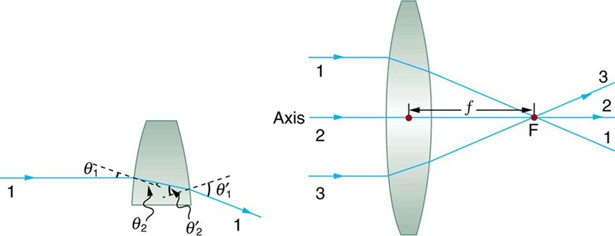
The lens in which light rays that enter it parallel to its axis cross one another at a single point on the opposite side with a converging effect is called converging lens.

Focal point f

The point at which the light rays cross is called the focal point F of the lens.

Focal length f size 12{f} {}

The distance from the center of the lens to its focal point is called focal length f size 12{f} {} .

Questions & Answers

what is biology
Hajah Reply
the study of living organisms and their interactions with one another and their environments
AI-Robot
what is biology
Victoria Reply
HOW CAN MAN ORGAN FUNCTION
Alfred Reply
the diagram of the digestive system
Assiatu Reply
allimentary cannel
Ogenrwot
How does twins formed
William Reply
They formed in two ways first when one sperm and one egg are splited by mitosis or two sperm and two eggs join together
Oluwatobi
what is genetics
Josephine Reply
Genetics is the study of heredity
Misack
how does twins formed?
Misack
What is manual
Hassan Reply
discuss biological phenomenon and provide pieces of evidence to show that it was responsible for the formation of eukaryotic organelles
Joseph Reply
what is biology
Yousuf Reply
the study of living organisms and their interactions with one another and their environment.
Wine
discuss the biological phenomenon and provide pieces of evidence to show that it was responsible for the formation of eukaryotic organelles in an essay form
Joseph Reply
what is the blood cells
Shaker Reply
list any five characteristics of the blood cells
Shaker
lack electricity and its more savely than electronic microscope because its naturally by using of light
Abdullahi Reply
advantage of electronic microscope is easily and clearly while disadvantage is dangerous because its electronic. advantage of light microscope is savely and naturally by sun while disadvantage is not easily,means its not sharp and not clear
Abdullahi
cell theory state that every organisms composed of one or more cell,cell is the basic unit of life
Abdullahi
is like gone fail us
DENG
cells is the basic structure and functions of all living things
Ramadan
What is classification
ISCONT Reply
is organisms that are similar into groups called tara
Yamosa
in what situation (s) would be the use of a scanning electron microscope be ideal and why?
Kenna Reply
A scanning electron microscope (SEM) is ideal for situations requiring high-resolution imaging of surfaces. It is commonly used in materials science, biology, and geology to examine the topography and composition of samples at a nanoscale level. SEM is particularly useful for studying fine details,
Hilary
cell is the building block of life.
Condoleezza Reply
Got questions? Join the online conversation and get instant answers!
Jobilize.com Reply
Practice Key Terms 8

Get Jobilize Job Search Mobile App in your pocket Now!

Get it on Google Play Download on the App Store Now


Source:  OpenStax, College physics for ap® courses. OpenStax CNX. Nov 04, 2016 Download for free at https://legacy.cnx.org/content/col11844/1.14
Google Play and the Google Play logo are trademarks of Google Inc.

Notification Switch

Would you like to follow the 'College physics for ap® courses' conversation and receive update notifications?

Ask