<< Chapter < Page Chapter >> Page >
Diagram of DNA replication. A small inset at the top shows a double strand of DNA separated in the center forming a bubble; the DNA is double stranded on either side of the bubble. The origin of replication is in the midway point of the bubble. On the top strand a solid arrow points to the left from the origin; this is the leading strand. On the right of the origin of replication are short arrows pointing to the left; this is the lagging strand. On the bottom strand a solid arrow pointing to the right from the origin is labeled leading strand and short arrows pointing to the right on the other side of the origin are labeled lagging strands. A larger image shows just the left half of the bubble. The double stranded DNA is no the far left and is labeled 5’ for the top strand and 3’ for the bottom strand. An enzyme to the very far left I is labeled topoisomerase/gyrase. At the point where the double stranded regions splits is a triangle shape labeled helicase. Next to that are smaller shapes labeled single-stranded binding proteins. The top strand shows continuous synthesis of the leading strand; this is shown as a solid arrow under the top strand. The arrow has a 5’ at the right end and a 3’ at the left end. The template strand at the top has a 3’ at the right and a 5’ at the left. At the end of the arrow (near where the DNA is newly being separated by the helicase) is DNA polymerase 3 and a sliding clamp that span both strands. The bottom strand of DNA has more components. Just after the single stranded binding proteins is RNA primase which attaches RNA primer (shown as a green arrow). Further down the lagging strand template is an existing RNA primer with DNA polymerase III and a sliding clamp spanning primer and the template strand. The polymerase is building a new strand of DNA from the left side (5’) to the right side (3’). Further to the right is a long piece made of RNA primer, then new DNA, then RNA primer, then new DNA all connected. Each of the DNA/RNA combinations are okazaki fragments made in the discontinuous synthesis of the lagging strand. DNA polymerase I is attached to the RNA primer in the center and is replacing it with DNA nucleotides. DNA ligase then binds the individual strands of new DNA together. This is shown in a close-up as two double helices that have all the correct letters in place, but one is missing a connection between two of the nucleotides (this is called a single-stranded gap). DNA ligase forms this last bond and the gap is sealed.
At the origin of replication, topoisomerase II relaxes the supercoiled chromosome. Two replication forks are formed by the opening of the double-stranded DNA at the origin, and helicase separates the DNA strands, which are coated by single-stranded binding proteins to keep the strands separated. DNA replication occurs in both directions. An RNA primer complementary to the parental strand is synthesized by RNA primase and is elongated by DNA polymerase III through the addition of nucleotides to the 3’-OH end. On the leading strand, DNA is synthesized continuously, whereas on the lagging strand, DNA is synthesized in short stretches called Okazaki fragments. RNA primers within the lagging strand are removed by the exonuclease activity of DNA polymerase I, and the Okazaki fragments are joined by DNA ligase.

Termination

Once the complete chromosome has been replicated, termination of DNA replication must occur. Although much is known about initiation of replication, less is known about the termination process. Following replication, the resulting complete circular genomes of prokaryotes are concatenated, meaning that the circular DNA chromosomes are interlocked and must be separated from each other. This is accomplished through the activity of bacterial topoisomerase IV, which introduces double-stranded breaks into DNA molecules, allowing them to separate from each other; the enzyme then reseals the circular chromosomes. The resolution of concatemers is an issue unique to prokaryotic DNA replication because of their circular chromosomes. Because both bacterial DNA gyrase and topoisomerase IV are distinct from their eukaryotic counterparts, these enzymes serve as targets for a class of antimicrobial drugs called quinolones .

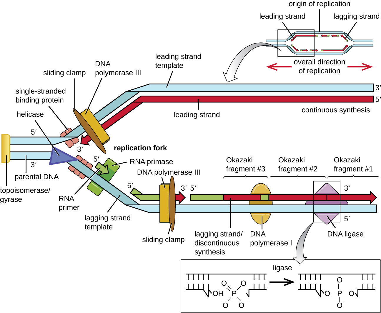
The Molecular Machinery Involved in Bacterial DNA Replication
Enzyme or Factor Function
DNA pol I Exonuclease activity removes RNA primer and replaces it with newly synthesized DNA
DNA pol III Main enzyme that adds nucleotides in the 5’ to 3’ direction
Helicase Opens the DNA helix by breaking hydrogen bonds between the nitrogenous bases
Ligase Seals the gaps between the Okazaki fragments on the lagging strand to create one continuous DNA strand
Primase Synthesizes RNA primers needed to start replication
Single-stranded binding proteins Bind to single-stranded DNA to prevent hydrogen bonding between DNA strands, reforming double-stranded DNA
Sliding clamp Helps hold DNA pol III in place when nucleotides are being added
Topoisomerase II (DNA gyrase) Relaxes supercoiled chromosome to make DNA more accessible for the initiation of replication; helps relieve the stress on DNA when unwinding, by causing breaks and then resealing the DNA
Topoisomerase IV Introduces single-stranded break into concatenated chromosomes to release them from each other, and then reseals the DNA
  • Which enzyme breaks the hydrogen bonds holding the two strands of DNA together so that replication can occur?
  • Is it the lagging strand or the leading strand that is synthesized in the direction toward the opening of the replication fork?
  • Which enzyme is responsible for removing the RNA primers in newly replicated bacterial DNA?

Questions & Answers

how does Neisseria cause meningitis
Nyibol Reply
what is microbiologist
Muhammad Reply
what is errata
Muhammad
is the branch of biology that deals with the study of microorganisms.
Ntefuni Reply
What is microbiology
Mercy Reply
studies of microbes
Louisiaste
when we takee the specimen which lumbar,spin,
Ziyad Reply
How bacteria create energy to survive?
Muhamad Reply
Bacteria doesn't produce energy they are dependent upon their substrate in case of lack of nutrients they are able to make spores which helps them to sustain in harsh environments
_Adnan
But not all bacteria make spores, l mean Eukaryotic cells have Mitochondria which acts as powerhouse for them, since bacteria don't have it, what is the substitution for it?
Muhamad
they make spores
Louisiaste
what is sporadic nd endemic, epidemic
Aminu Reply
the significance of food webs for disease transmission
Abreham
food webs brings about an infection as an individual depends on number of diseased foods or carriers dully.
Mark
explain assimilatory nitrate reduction
Esinniobiwa Reply
Assimilatory nitrate reduction is a process that occurs in some microorganisms, such as bacteria and archaea, in which nitrate (NO3-) is reduced to nitrite (NO2-), and then further reduced to ammonia (NH3).
Elkana
This process is called assimilatory nitrate reduction because the nitrogen that is produced is incorporated in the cells of microorganisms where it can be used in the synthesis of amino acids and other nitrogen products
Elkana
Examples of thermophilic organisms
Shu Reply
Give Examples of thermophilic organisms
Shu
advantages of normal Flora to the host
Micheal Reply
Prevent foreign microbes to the host
Abubakar
they provide healthier benefits to their hosts
ayesha
They are friends to host only when Host immune system is strong and become enemies when the host immune system is weakened . very bad relationship!
Mark
what is cell
faisal Reply
cell is the smallest unit of life
Fauziya
cell is the smallest unit of life
Akanni
ok
Innocent
cell is the structural and functional unit of life
Hasan
is the fundamental units of Life
Musa
what are emergency diseases
Micheal Reply
There are nothing like emergency disease but there are some common medical emergency which can occur simultaneously like Bleeding,heart attack,Breathing difficulties,severe pain heart stock.Hope you will get my point .Have a nice day ❣️
_Adnan
define infection ,prevention and control
Innocent
I think infection prevention and control is the avoidance of all things we do that gives out break of infections and promotion of health practices that promote life
Lubega
Heyy Lubega hussein where are u from?
_Adnan
en français
Adama
which site have a normal flora
ESTHER Reply
Many sites of the body have it Skin Nasal cavity Oral cavity Gastro intestinal tract
Safaa
skin
Asiina
skin,Oral,Nasal,GIt
Sadik
How can Commensal can Bacteria change into pathogen?
Sadik
How can Commensal Bacteria change into pathogen?
Sadik
all
Tesfaye
by fussion
Asiina
what are the advantages of normal Flora to the host
Micheal
what are the ways of control and prevention of nosocomial infection in the hospital
Micheal
what is inflammation
Shelly Reply
part of a tissue or an organ being wounded or bruised.
Wilfred
what term is used to name and classify microorganisms?
Micheal Reply
Binomial nomenclature
adeolu

Get Jobilize Job Search Mobile App in your pocket Now!

Get it on Google Play Download on the App Store Now


Source:  OpenStax, Microbiology. OpenStax CNX. Nov 01, 2016 Download for free at http://cnx.org/content/col12087/1.4
Google Play and the Google Play logo are trademarks of Google Inc.

Notification Switch

Would you like to follow the 'Microbiology' conversation and receive update notifications?

Ask