<< Chapter < Page Chapter >> Page >
a) phot of worms filling the intestines. B) photo of a large handful of worms. C) photo of a circle in a thicker circle. The outer circle is about 60 micrometers.
(a) Adult Ascaris lumbricoides roundworms can cause intestinal blockage. (b) This mass of A. lumbricoides worms was excreted by a child. (c) A micrograph of a fertilized egg of A. lumbricoides. Fertilized eggs can be distinguished from unfertilized eggs because they are round rather than elongated and have a thicker cell wall. (credit a: modification of work by South African Medical Research Council; credit b: modification of work by James Gathany, Centers for Disease Control and Prevention; credit c: modification of work by Centers for Disease Control and Prevention)
  • Describe the route by which A. lumbricoides reaches the host’s intestines as an adult worm.

Hookworm

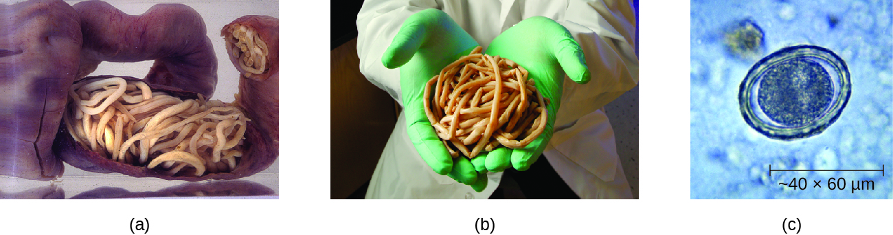
Two species of nematode worms are associated with hookworm infection . Both species are found in the Americas, Africa, and Asia. Necator americanus is found predominantly in the United States and Australia. Another species, Ancylostoma doudenale , is found in southern Europe, North Africa, the Middle East, and Asia.

The eggs of these species develop into larvae in soil contaminated by dog or cat feces. These larvae can penetrate the skin. After traveling through the venous circulation, they reach the lungs. When they are coughed up, they are then swallowed and can enter the intestine and develop into mature adults. At this stage, they attach to the wall of the intestine, where they feed on blood and can potentially cause anemia. Signs and symptoms include cough, an itchy rash, loss of appetite, abdominal pain, and diarrhea. In children, hookworms can affect physical and cognitive growth.

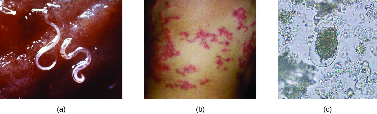
Some hookworm species, such as Ancylostoma braziliense that is commonly found in animals such as cats and dogs, can penetrate human skin and migrate, causing cutaneous larva migrans , a skin disease caused by the larvae of hookworms. As they move across the skin, in the subcutaneous tissue, pruritic tracks appear ( [link] ).

The infection is diagnosed using microscopic examination of the stool, allowing for observation of eggs in the feces. Medications such as albendazole , mebendazole , and pyrantel pamoate are used as needed to treat systemic infection. In addition to systemic medication for symptoms associated with cutaneous larva migrans, topical thiabendazole is applied to the affected areas.

a) photo of a cler worm attached to tissue. B) photo of red lines in the skin. c) Micrograph of an oval structure.
(a) This animal hookworm, Ancylostoma caninum , is attached to the intestinal wall. (b) The tracks of hookworms are visible in this individual with cutaneous larva migrans. (c) This micrograph shows the microscopic egg of a hookworm. (credit a, c: modification of work by Centers for Disease Control and Prevention)

Strongyloidiasis

Strongyloidiasis is generally caused by Strongyloides stercoralis , a soil-transmitted helminth with both free-living and parasitic forms. In the parasitic form, the larvae of these nematodes generally penetrate the body through the skin, especially through bare feet, although transmission through organ transplantation or at facilities like day-care centers can also occur. When excreted in the stool, larvae can become free-living adults rather than developing into the parasitic form. These free-living worms reproduce, laying eggs that hatch into larvae that can develop into the parasitic form. In the parasitic life cycle, infective larvae enter the skin, generally through the feet. The larvae reach the circulatory system, which allows them to travel to the alveolar spaces of the lungs. They are transported to the pharynx where, like many other helminths, the infected patient coughs them up and swallows them again so that they return to the intestine . Once they reach the intestine, females live in the epithelium and produce eggs that develop asexually, unlike the free-living forms, which use sexual reproduction. The larvae may be excreted in the stool or can reinfect the host by entering the tissue of the intestines and skin around the anus, which can lead to chronic infections.

Questions & Answers

what is phylogeny
Odigie Reply
evolutionary history and relationship of an organism or group of organisms
AI-Robot
ok
Deng
what is biology
Hajah Reply
the study of living organisms and their interactions with one another and their environments
AI-Robot
what is biology
Victoria Reply
HOW CAN MAN ORGAN FUNCTION
Alfred Reply
the diagram of the digestive system
Assiatu Reply
allimentary cannel
Ogenrwot
How does twins formed
William Reply
They formed in two ways first when one sperm and one egg are splited by mitosis or two sperm and two eggs join together
Oluwatobi
what is genetics
Josephine Reply
Genetics is the study of heredity
Misack
how does twins formed?
Misack
What is manual
Hassan Reply
discuss biological phenomenon and provide pieces of evidence to show that it was responsible for the formation of eukaryotic organelles
Joseph Reply
what is biology
Yousuf Reply
the study of living organisms and their interactions with one another and their environment.
Wine
discuss the biological phenomenon and provide pieces of evidence to show that it was responsible for the formation of eukaryotic organelles in an essay form
Joseph Reply
what is the blood cells
Shaker Reply
list any five characteristics of the blood cells
Shaker
lack electricity and its more savely than electronic microscope because its naturally by using of light
Abdullahi Reply
advantage of electronic microscope is easily and clearly while disadvantage is dangerous because its electronic. advantage of light microscope is savely and naturally by sun while disadvantage is not easily,means its not sharp and not clear
Abdullahi
cell theory state that every organisms composed of one or more cell,cell is the basic unit of life
Abdullahi
is like gone fail us
DENG
cells is the basic structure and functions of all living things
Ramadan
What is classification
ISCONT Reply
is organisms that are similar into groups called tara
Yamosa
in what situation (s) would be the use of a scanning electron microscope be ideal and why?
Kenna Reply
A scanning electron microscope (SEM) is ideal for situations requiring high-resolution imaging of surfaces. It is commonly used in materials science, biology, and geology to examine the topography and composition of samples at a nanoscale level. SEM is particularly useful for studying fine details,
Hilary
Got questions? Join the online conversation and get instant answers!
Jobilize.com Reply

Get Jobilize Job Search Mobile App in your pocket Now!

Get it on Google Play Download on the App Store Now


Source:  OpenStax, Microbiology. OpenStax CNX. Nov 01, 2016 Download for free at http://cnx.org/content/col12087/1.4
Google Play and the Google Play logo are trademarks of Google Inc.

Notification Switch

Would you like to follow the 'Microbiology' conversation and receive update notifications?

Ask