<< Chapter < Page Chapter >> Page >
A pregnancy test stick with 2 red lines; one is labeled test line and the other is labeled control line. A key on the stick states that 2 lines means pregnant and 1 line means not pregnant
A lateral flow test detecting pregnancy-related hormones in urine. The control stripe verifies the validity of the test and the test line determines the presence of pregnancy-related hormones in the urine. (credit: modification of work by Klaus Hoffmeier)
A diagram showing lateral flow test of a urine sample. The top panel shows a positive test and the bottom shows a negative test. In both a urine sample is added; in the positive sample there are antigens present. The label reads hCG urine sample is applied to absorbent sample pad. In both cases the urine sample flows across the test area. The first region is a mix area that contains hCG-second antibody-AuNPs (hCG-GC). These then flow across the hCG strip in both samples. Next, we reach the test line. Here there are antibodies that bind the antigens in the positive sample but bind nothing in the negative sample. In the positive sample the hCG-GC also bind to the antigen, forming an antibody sandwich around the antigen. The presence of the hCG-GC causes a color change here in the positive sample but not in the negative sample.  Finally, the control line contains antibodies that bind the hCG-GC directly; so these bind in both the positive and negative samples. The control line turns a color in both.
Immunochromatographic assays, or lateral flow tests, allow the testing of antigen in a dilute solution. As the fluid flows through the test strip, it rehydrates the reagents. Antibodies conjugated to small particles bind the antigen in the first stripe and then flow onto the second stripe where they are bound by a second, fixed antibody. This produces a line of color, depending on the color of the beads. The third, control stripe binds beads as well to indicate that the test is working properly. (credit: modification of work by Yeh CH, Zhao ZQ, Shen PL, Lin YC)
  • What physical process does the lateral flow method require to function?
  • Explain the purpose of the third strip in a lateral flow assay.

[link] compares some of the key mechanisms and examples of some of the EIAs discussed in this section as well as immunoblots, which were discussed in Detecting Antigen-Antibody Complexes .

Immunoblots&Enzyme Immunoassays
Type of Assay Mechanism Specific Procedures Examples
Immunoblots Uses enzyme-antibody conjugates to identify specific proteins that have been transferred to an absorbent membrane Western blot: Detects the presence of a particular protein Detecting the presence of HIV peptides (or peptides from other infectious agents) in patient sera
Immunostaining Uses enzyme-antibody conjugates to stain specific molecules on or in cells Immunohistochemistry: Used to stain specific cells in a tissue Stain for presence of CD8 cells in host tissue
Enzyme-linked immunosorbent assay (ELISA) Uses enzyme-antibody conjugates to quantify target molecules Direct ELISA: Uses a single antibody to detect the presence of an antigen Detection of HIV antigen p24 up to one month after being infected
Indirect ELISA: Measures the amount of antibody produced against an antigen Detection of HIV antibodies in serum
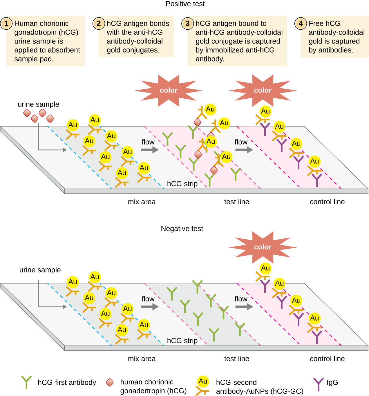
Immunochromatographic (lateral flow) assays Techniques use the capture of flowing, color-labeled antigen-antibody complexes by fixed antibody for disease diagnosis Sandwich ELISA: Measures the amount of antigen bound by the antibody Detection of antibodies for various pathogens in patient sera (e.g., rapid strep, malaria dipstick)
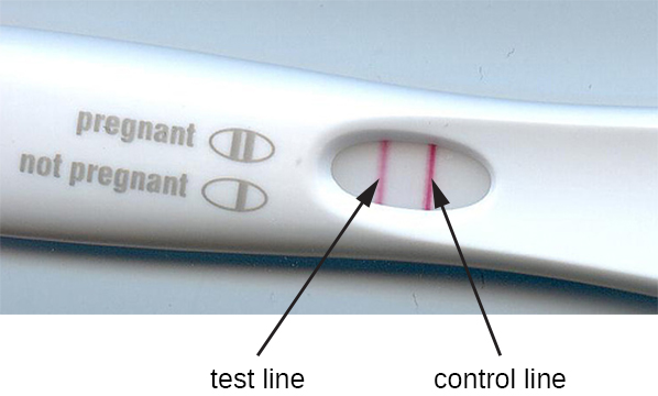
Pregnancy test detecting human chorionic gonadotrophin in urine

Part 3

Although the indirect ELISA for HIV is a sensitive assay, there are several complicating considerations. First, if an infected person is tested too soon after becoming infected, the test can yield false-negative results. The seroconversion window is generally about three weeks, but in some cases, it can be more than two months.

In addition to false negatives, false positives can also occur, usually due to previous infections with other viruses that induce cross-reacting antibodies. The false-positive rate depends on the particular brand of test used, but 0.5% is not unusual. Thomas, Justin G., Victor Jaffe, Judith Shaffer, and Jose Abreu, “HIV Testing: US Recommendations 2014,” Osteopathic Family Physician 6, no. 6 (2014). Because of the possibility of a false positive, all positive tests are followed up with a confirmatory test. This confirmatory test is often an immunoblot (western blot) in which HIV peptides from the patient’s blood are identified using an HIV-specific mAb-enzyme conjugate. A positive western blot would confirm an HIV infection and a negative blot would confirm the absence of HIV despite the positive ELISA.

Unfortunately, western blots for HIV antigens often yield indeterminant results, in which case, they neither confirm nor invalidate the results of the indirect ELISA. In fact, the rate of indeterminants can be 10–49% (which is why, combined with their cost, western blots are not used for screening). Similar to the indirect ELISA, an indeterminant western blot can occur because of cross-reactivity or previous viral infections, vaccinations, or autoimmune diseases.

  • Of the 1300 patients being tested, how many false-positive ELISA tests would be expected?
  • Of the false positives, how many indeterminant western blots could be expected?
  • How would the hospital address any cases in which a patient’s western blot was indeterminant?

Jump to the previous Clinical Focus box. Jump to the next Clinical Focus box.

Key concepts and summary

  • Enzyme immunoassays (EIA) are used to visualize and quantify antigens. They use an antibody conjugated to an enzyme to bind the antigen, and the enzyme converts a substrate into an observable end product. The substrate may be either a chromogen or a fluorogen.
  • Immunostaining is an EIA technique for visualizing cells in a tissue ( immunohistochemistry ) or examining intracellular structures ( immunocytochemistry ).
  • Direct ELISA is used to quantify an antigen in solution. The primary antibody captures the antigen, and the secondary antibody delivers an enzyme. Production of end product from the chromogenic substrate is directly proportional to the amount of captured antigen.
  • Indirect ELISA is used to detect antibodies in patient serum by attaching antigen to the well of a microtiter plate, allowing the patient (primary) antibody to bind the antigen and an enzyme-conjugated secondary antibody to detect the primary antibody.
  • Immunofiltration and immunochromatographic assays are used in lateral flow tests , which can be used to diagnose pregnancy and various diseases by detecting color-labeled antigen-antibody complexes in urine or other fluid samples

Fill in the blank

To detect antibodies against bacteria in the bloodstream using an EIA, we would run a(n) ________, which we would start by attaching antigen from the bacteria to the wells of a microtiter plate.

indirect ELISA

Got questions? Get instant answers now!

Short answer

Why is it important in a sandwich ELISA that the antigen has multiple epitopes? And why might it be advantageous to use polyclonal antisera rather than mAb in this assay?

Got questions? Get instant answers now!

The pregnancy test strip detects the presence of human chorionic gonadotrophin in urine. This hormone is initially produced by the fetus and later by the placenta. Why is the test strip preferred for this test rather than using either a direct or indirect ELISA with their more quantifiable results?

Got questions? Get instant answers now!

Questions & Answers

what is biology
Hajah Reply
the study of living organisms and their interactions with one another and their environments
AI-Robot
what is biology
Victoria Reply
HOW CAN MAN ORGAN FUNCTION
Alfred Reply
the diagram of the digestive system
Assiatu Reply
allimentary cannel
Ogenrwot
How does twins formed
William Reply
They formed in two ways first when one sperm and one egg are splited by mitosis or two sperm and two eggs join together
Oluwatobi
what is genetics
Josephine Reply
Genetics is the study of heredity
Misack
how does twins formed?
Misack
What is manual
Hassan Reply
discuss biological phenomenon and provide pieces of evidence to show that it was responsible for the formation of eukaryotic organelles
Joseph Reply
what is biology
Yousuf Reply
the study of living organisms and their interactions with one another and their environment.
Wine
discuss the biological phenomenon and provide pieces of evidence to show that it was responsible for the formation of eukaryotic organelles in an essay form
Joseph Reply
what is the blood cells
Shaker Reply
list any five characteristics of the blood cells
Shaker
lack electricity and its more savely than electronic microscope because its naturally by using of light
Abdullahi Reply
advantage of electronic microscope is easily and clearly while disadvantage is dangerous because its electronic. advantage of light microscope is savely and naturally by sun while disadvantage is not easily,means its not sharp and not clear
Abdullahi
cell theory state that every organisms composed of one or more cell,cell is the basic unit of life
Abdullahi
is like gone fail us
DENG
cells is the basic structure and functions of all living things
Ramadan
What is classification
ISCONT Reply
is organisms that are similar into groups called tara
Yamosa
in what situation (s) would be the use of a scanning electron microscope be ideal and why?
Kenna Reply
A scanning electron microscope (SEM) is ideal for situations requiring high-resolution imaging of surfaces. It is commonly used in materials science, biology, and geology to examine the topography and composition of samples at a nanoscale level. SEM is particularly useful for studying fine details,
Hilary
cell is the building block of life.
Condoleezza Reply
Got questions? Join the online conversation and get instant answers!
Jobilize.com Reply

Get Jobilize Job Search Mobile App in your pocket Now!

Get it on Google Play Download on the App Store Now


Source:  OpenStax, Microbiology. OpenStax CNX. Nov 01, 2016 Download for free at http://cnx.org/content/col12087/1.4
Google Play and the Google Play logo are trademarks of Google Inc.

Notification Switch

Would you like to follow the 'Microbiology' conversation and receive update notifications?

Ask