<< Chapter < Page Chapter >> Page >

Learning objectives

  • Compare direct and indirect agglutination
  • Identify various uses of hemagglutination in the diagnosis of disease
  • Explain how blood types are determined
  • Explain the steps used to cross-match blood to be used in a transfusion

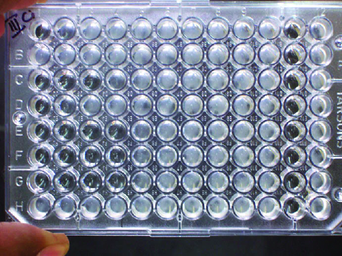
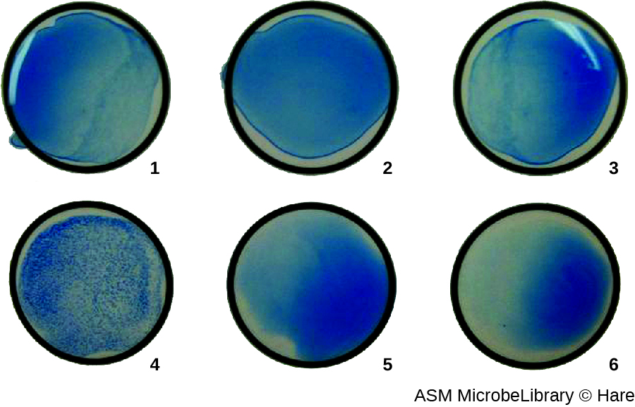
In addition to causing precipitation of soluble molecules and flocculation of molecules in suspension, antibodies can also clump together cells or particles (e.g., antigen-coated latex beads) in a process called agglutination ( [link] ). Agglutination can be used as an indicator of the presence of antibodies against bacteria or red blood cells. Agglutination assays are usually quick and easy to perform on a glass slide or microtiter plate ( [link] ). Microtiter plates have an array of wells to hold small volumes of reagents and to observe reactions (e.g., agglutination) either visually or using a specially designed spectrophotometer. The wells come in many different sizes for assays involving different volumes of reagents.

A plastic plate about the size of a hand with wells in a grid. The top is labeled 1-12 and the side is labeled A-H.
Microtiter plates are used for conducting numerous reactions simultaneously in an array of wells. (credit: modification of work by “Microrao”/Wikimedia)

Agglutination of bacteria and viruses

The use of agglutination tests to identify streptococcal bacteria was developed in the 1920s by Rebecca Lancefield working with her colleagues A.R. Dochez and Oswald Avery . Lancefield, Rebecca C., “The Antigenic Complex of Streptococcus haemoliticus . I. Demonstration of a Type-Specific Substance in Extracts of Streptococcus haemolyticus ,” The Journal of Experimental Medicine 47, no. 1 (1928): 91-103. She used antibodies to identify M protein , a virulence factor on streptococci that is necessary for the bacteria’s ability to cause strep throat. Production of antibodies against M protein is crucial in mounting a protective response against the bacteria.

Lancefield used antisera to show that different strains of the same species of streptococci express different versions of M protein, which explains why children can come down with strep throat repeatedly. Lancefield classified beta-hemolytic streptococci into many groups based on antigenic differences in group-specific polysaccharides located in the bacterial cell wall. The strains are called serovars because they are differentiated using antisera. Identifying the serovars present in a disease outbreak is important because some serovars may cause more severe disease than others.

The method developed by Lancefield is a direct agglutination assay , since the bacterial cells themselves agglutinate. A similar strategy is more commonly used today when identifying serovars of bacteria and viruses; however, to improve visualization of the agglutination, the antibodies may be attached to inert latex beads . This technique is called an indirect agglutination assay (or latex fixation assay ), because the agglutination of the beads is a marker for antibody binding to some other antigen ( [link] ). Indirect assays can be used to detect the presence of either antibodies or specific antigens.

Photo of 6 wells. Well 4 has blue spots. All the other wells have a clear blue color.
Antibodies against six different serovars of Group A strep were attached to latex beads. Each of the six antibody preparations was mixed with bacteria isolated from a patient. The tiny clumps seen in well 4 are indicative of agglutination, which is absent from all other wells. This indicates that the serovar associated with well 4 is present in the patient sample. (credit: modification of work by American Society for Microbiology)

Questions & Answers

what is phylogeny
Odigie Reply
evolutionary history and relationship of an organism or group of organisms
AI-Robot
ok
Deng
what is biology
Hajah Reply
the study of living organisms and their interactions with one another and their environments
AI-Robot
what is biology
Victoria Reply
HOW CAN MAN ORGAN FUNCTION
Alfred Reply
the diagram of the digestive system
Assiatu Reply
allimentary cannel
Ogenrwot
How does twins formed
William Reply
They formed in two ways first when one sperm and one egg are splited by mitosis or two sperm and two eggs join together
Oluwatobi
what is genetics
Josephine Reply
Genetics is the study of heredity
Misack
how does twins formed?
Misack
What is manual
Hassan Reply
discuss biological phenomenon and provide pieces of evidence to show that it was responsible for the formation of eukaryotic organelles
Joseph Reply
what is biology
Yousuf Reply
the study of living organisms and their interactions with one another and their environment.
Wine
discuss the biological phenomenon and provide pieces of evidence to show that it was responsible for the formation of eukaryotic organelles in an essay form
Joseph Reply
what is the blood cells
Shaker Reply
list any five characteristics of the blood cells
Shaker
lack electricity and its more savely than electronic microscope because its naturally by using of light
Abdullahi Reply
advantage of electronic microscope is easily and clearly while disadvantage is dangerous because its electronic. advantage of light microscope is savely and naturally by sun while disadvantage is not easily,means its not sharp and not clear
Abdullahi
cell theory state that every organisms composed of one or more cell,cell is the basic unit of life
Abdullahi
is like gone fail us
DENG
cells is the basic structure and functions of all living things
Ramadan
What is classification
ISCONT Reply
is organisms that are similar into groups called tara
Yamosa
in what situation (s) would be the use of a scanning electron microscope be ideal and why?
Kenna Reply
A scanning electron microscope (SEM) is ideal for situations requiring high-resolution imaging of surfaces. It is commonly used in materials science, biology, and geology to examine the topography and composition of samples at a nanoscale level. SEM is particularly useful for studying fine details,
Hilary
Got questions? Join the online conversation and get instant answers!
Jobilize.com Reply

Get Jobilize Job Search Mobile App in your pocket Now!

Get it on Google Play Download on the App Store Now


Source:  OpenStax, Microbiology. OpenStax CNX. Nov 01, 2016 Download for free at http://cnx.org/content/col12087/1.4
Google Play and the Google Play logo are trademarks of Google Inc.

Notification Switch

Would you like to follow the 'Microbiology' conversation and receive update notifications?

Ask