<< Chapter < Page Chapter >> Page >

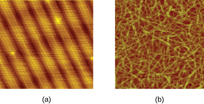
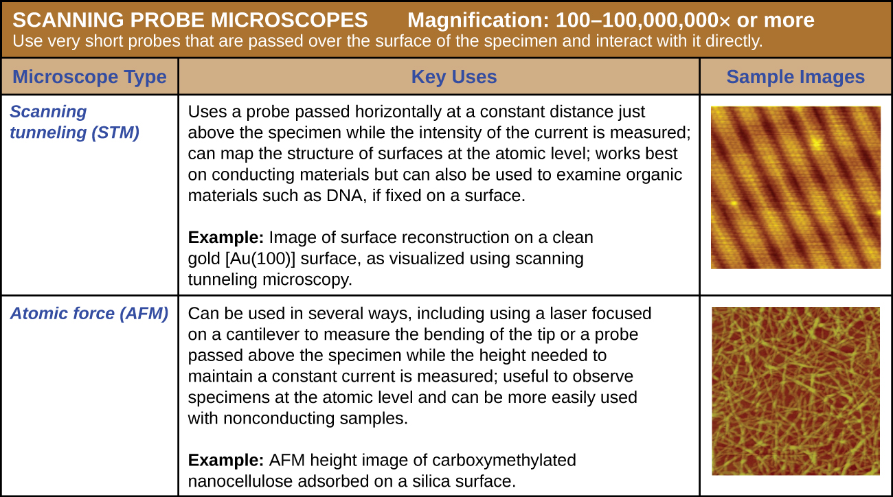
There are two types of scanning probe microscope: the scanning tunneling microscope (STM) and the atomic force microscope (AFM) . An STM uses a probe that is passed just above the specimen as a constant voltage bias creates the potential for an electric current between the probe and the specimen. This current occurs via quantum tunneling of electrons between the probe and the specimen, and the intensity of the current is dependent upon the distance between the probe and the specimen. The probe is moved horizontally above the surface and the intensity of the current is measured. Scanning tunneling microscopy can effectively map the structure of surfaces at a resolution at which individual atoms can be detected.

Similar to an STM, AFMs have a thin probe that is passed just above the specimen. However, rather than measuring variations in the current at a constant height above the specimen, an AFM establishes a constant current and measures variations in the height of the probe tip as it passes over the specimen. As the probe tip is passed over the specimen, forces between the atoms (van der Waals forces, capillary forces, chemical bonding, electrostatic forces, and others) cause it to move up and down. Deflection of the probe tip is determined and measured using Hooke’s law of elasticity , and this information is used to construct images of the surface of the specimen with resolution at the atomic level ( [link] ).

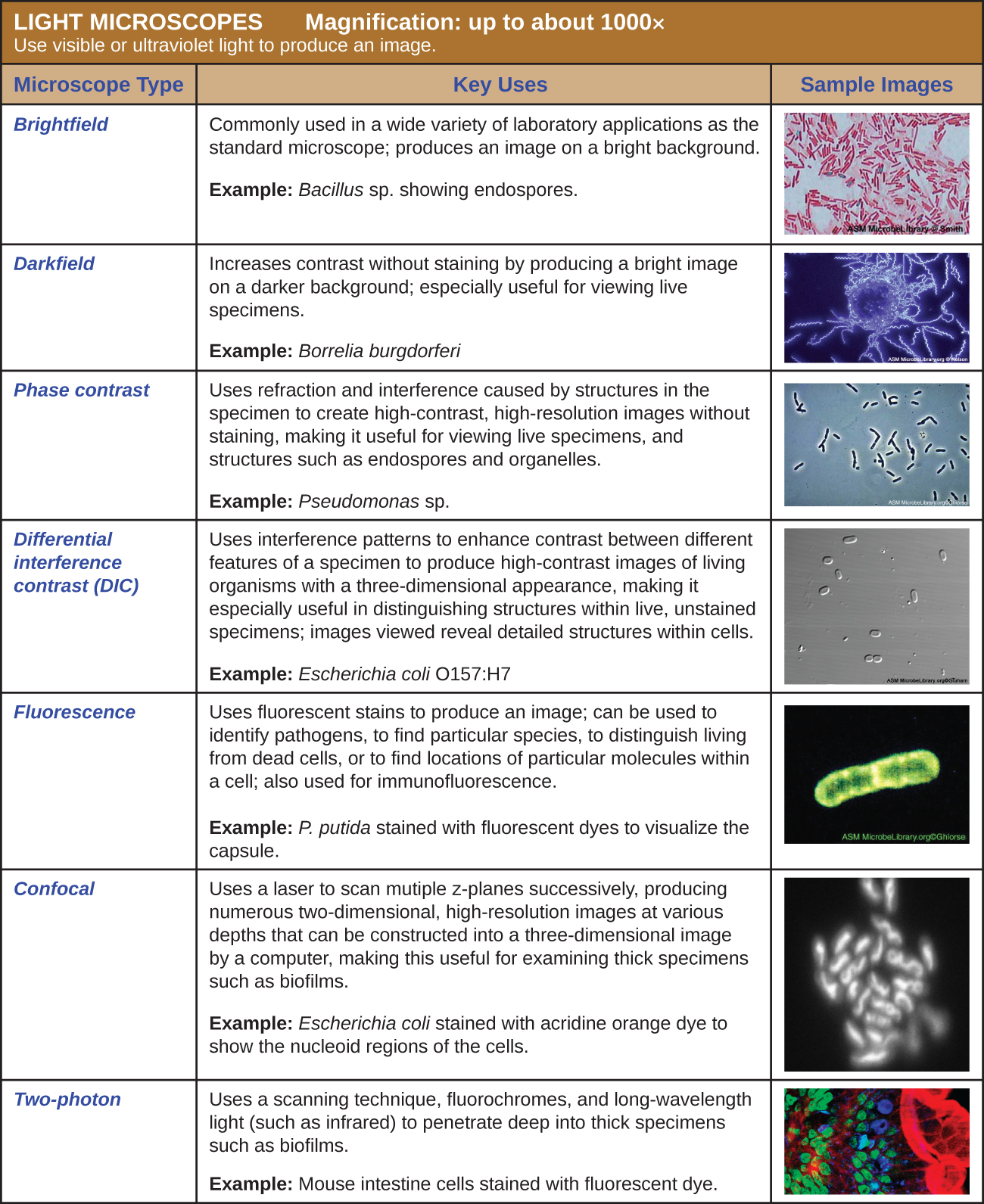
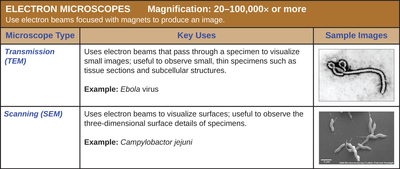
[link] , [link] , and [link] summarize the microscopy techniques for light microscopes, electron microscopes, and scanning probe microscopes, respectively.

Micrograph a shows circle arranged in repeating rows. Micrograph b shows long strands in a pile.
STMs and AFMs allow us to view images at the atomic level. (a) This STM image of a pure gold surface shows individual atoms of gold arranged in columns. (b) This AFM image shows long, strand-like molecules of nanocellulose, a laboratory-created substance derived from plant fibers. (credit a: modification of work by “Erwinrossen”/Wikimedia Commons)
  • Which has higher magnification, a light microscope or a scanning probe microscope?
  • Name one advantage and one limitation of scanning probe microscopy.
A table of light microscope types. These use visible or ultraviolet light to produce an image. Magnification: up to about 1000x. Brightfield microscopes are commonly used in a wide variety of laboratory applications as the standard microscope and produce an image on a bright background. The sample image of Bacillus sp. shows red rods on a clear background; small green dots in the red cells indicate endospores. Darkfield microscopes increase contrast without staining by producing a bright image on a dark background. These are especially useful for viewing live specimens. The sample image (Borrelia burgdorferi) shows bright spirals on a dark background. Phase contrast microscopes use refraction and interference caused by structures in the specimen to create high-contrast, high-resolution images without staining, making it useful for viewing live specimens and structures such as endospores and organelles. The sample image (Pseudomonas sp.) shows dark rods with a bright halo. Differential interference contrast (DIC) uses interference patters to enhance contrast between different features of a specimen to produce high-contrast images of living organisms with a three-dimensional appearance, making it especially useful in distinguishing structures within live, unstained specimens. Images viewed reveal detailed structures within cells. The sample image (Escherichia coli 0157:H7) shows small three-dimensional ovals. Fluorescence uses fluorescent stains to produce an image. Fluorescent microscopes can be used to identify pathogens, to find particular species, to distinguish living from dead, or to find location of particular molecules within a cell; also used for immunofluorescence. The sample image (Pseuodomonas putida stained with fluorescent dyes to visualize capsule) shows a green rod on a black background. Confocal microscopes use a laser to scan multiple z-planes successively, producing numerous two-dimensional, high-resolution images at various depths that can be constructed into a three-dimensional image by a computer, making this useful for examining thick specimens such as biofilms. The sample image (mouse intestine cells stained with fluorescent dye) shows cells of various colors on a dark background.
(credit “Brightfield”: modification of work by American Society for Microbiology; credit “Darkfield”: modification of work by American Society for Microbiology; credit “Phase contrast”: modification of work by American Society for Microbiology; credit “DIC”: modification of work by American Society for Microbiology; credit “Fluorescence”: modification of work by American Society for Microbiology; credit “Confocal”: modification of work by American Society for Microbiology; credit “Two-photon”: modification of work by Alberto Diaspro, Paolo Bianchini, Giuseppe Vicidomini, Mario Faretta, Paola Ramoino, Cesare Usai)
Table of electron microscopes which use electron beams focused with magnets to produce an image. Magnification: 20–100,000 x or more. Transmission electron microscopes (TEM) use electron means that pass through a specimen to visual small images; useful to observe small, thin specimens such as tissue sections and subcellular structures. The sample image (Ebola virus) shows a tube shaped into a letter d at one end. Scanning electron microscopes (SEM) use electron beams to visualize surfaces; useful to observe the three-dimensional surface details of specimens. The sample image (Campylobactor jejuni) shows thick three-dimensional spirals.
(credit “TEM”: modification of work by American Society for Microbiology; credit “SEM”: modification of work by American Society for Microbiology)
A table of scanning probe microscopes with very sharp probes that are passed over the surface of the specimen and interact with it directly. Magnification: 100–100,000,000x or more. A scanning tunneling microscope (STM) uses a probe passed horizontally at a constant distance just above the specimen while the intensity of the current is measured; can map the structure of surfaces at the atomic level; works best on conducting materials but can also be used to examine organic materials such as DNA if fixed on a surface. The sample image (of a gold surface) shows small circles in repeating rows. Atomic force microscopes (AFM) are used in several ways, including using a laser focused on a cantilever to measure the bending of the tip or a probe passed above the specimen while the height needs to maintain a constant current is measured; useful to observe specimens at the atomic level and can be more easily used with nonconducting samples. The sample image (carboxymethylated nanocellulse absorbed on a silica surface) shows long strands throughout.

Key concepts and summary

  • Numerous types of microscopes use various technologies to generate micrographs. Most are useful for a particular type of specimen or application.
  • Light microscopy uses lenses to focus light on a specimen to produce an image. Commonly used light microscopes include brightfield , darkfield , phase-contrast , differential interference contrast , fluorescence , confocal , and two-photon microscopes.
  • Electron microscopy focuses electrons on the specimen using magnets, producing much greater magnification than light microscopy. The transmission electron microscope (TEM) and scanning electron microscope (SEM) are two common forms.
  • Scanning probe microscopy produces images of even greater magnification by measuring feedback from sharp probes that interact with the specimen. Probe microscopes include the scanning tunneling microscope (STM) and the atomic force microscope (AFM) .

Fill in the blank

Chromophores that absorb and then emit light are called __________.

fluorochromes

Got questions? Get instant answers now!

In a(n) _______ microscope, a probe located just above the specimen moves up and down in response to forces between the atoms and the tip of the probe.

atomic force microscope

Got questions? Get instant answers now!

What is the total magnification of a specimen that is being viewed with a standard ocular lens and a 40⨯ objective lens?

400⨯

Got questions? Get instant answers now!

Short answer

What is the function of the condenser in a brightfield microscope?

Got questions? Get instant answers now!

Art connection

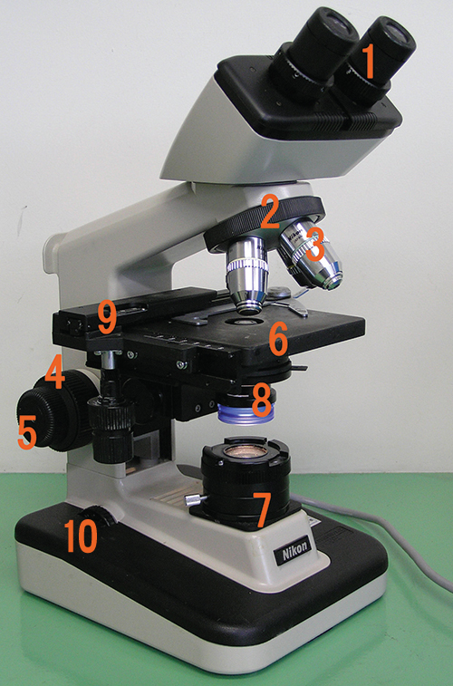
Label each component of the brightfield microscope.

A photo of a microscope is shown. The base contains a light source (#7) and a knob (#10). Attached at one end of the base is an arm with a projection to hold the specimen (#9). The center of #9 has an opening to allow light through. Below this opening are #6 & #8 (6 is above 8). Above this opening are four lenses (#3) attached to #2. Above the objective is #1. Attached to the bottom of the stage are two knobs (#9). On the arm below the stage are two knobs: #4 is larger than #5.
Got questions? Get instant answers now!

Questions & Answers

how does Neisseria cause meningitis
Nyibol Reply
what is microbiologist
Muhammad Reply
what is errata
Muhammad
is the branch of biology that deals with the study of microorganisms.
Ntefuni Reply
What is microbiology
Mercy Reply
studies of microbes
Louisiaste
when we takee the specimen which lumbar,spin,
Ziyad Reply
How bacteria create energy to survive?
Muhamad Reply
Bacteria doesn't produce energy they are dependent upon their substrate in case of lack of nutrients they are able to make spores which helps them to sustain in harsh environments
_Adnan
But not all bacteria make spores, l mean Eukaryotic cells have Mitochondria which acts as powerhouse for them, since bacteria don't have it, what is the substitution for it?
Muhamad
they make spores
Louisiaste
what is sporadic nd endemic, epidemic
Aminu Reply
the significance of food webs for disease transmission
Abreham
food webs brings about an infection as an individual depends on number of diseased foods or carriers dully.
Mark
explain assimilatory nitrate reduction
Esinniobiwa Reply
Assimilatory nitrate reduction is a process that occurs in some microorganisms, such as bacteria and archaea, in which nitrate (NO3-) is reduced to nitrite (NO2-), and then further reduced to ammonia (NH3).
Elkana
This process is called assimilatory nitrate reduction because the nitrogen that is produced is incorporated in the cells of microorganisms where it can be used in the synthesis of amino acids and other nitrogen products
Elkana
Examples of thermophilic organisms
Shu Reply
Give Examples of thermophilic organisms
Shu
advantages of normal Flora to the host
Micheal Reply
Prevent foreign microbes to the host
Abubakar
they provide healthier benefits to their hosts
ayesha
They are friends to host only when Host immune system is strong and become enemies when the host immune system is weakened . very bad relationship!
Mark
what is cell
faisal Reply
cell is the smallest unit of life
Fauziya
cell is the smallest unit of life
Akanni
ok
Innocent
cell is the structural and functional unit of life
Hasan
is the fundamental units of Life
Musa
what are emergency diseases
Micheal Reply
There are nothing like emergency disease but there are some common medical emergency which can occur simultaneously like Bleeding,heart attack,Breathing difficulties,severe pain heart stock.Hope you will get my point .Have a nice day ❣️
_Adnan
define infection ,prevention and control
Innocent
I think infection prevention and control is the avoidance of all things we do that gives out break of infections and promotion of health practices that promote life
Lubega
Heyy Lubega hussein where are u from?
_Adnan
en français
Adama
which site have a normal flora
ESTHER Reply
Many sites of the body have it Skin Nasal cavity Oral cavity Gastro intestinal tract
Safaa
skin
Asiina
skin,Oral,Nasal,GIt
Sadik
How can Commensal can Bacteria change into pathogen?
Sadik
How can Commensal Bacteria change into pathogen?
Sadik
all
Tesfaye
by fussion
Asiina
what are the advantages of normal Flora to the host
Micheal
what are the ways of control and prevention of nosocomial infection in the hospital
Micheal
what is inflammation
Shelly Reply
part of a tissue or an organ being wounded or bruised.
Wilfred
what term is used to name and classify microorganisms?
Micheal Reply
Binomial nomenclature
adeolu

Get Jobilize Job Search Mobile App in your pocket Now!

Get it on Google Play Download on the App Store Now


Source:  OpenStax, Microbiology. OpenStax CNX. Nov 01, 2016 Download for free at http://cnx.org/content/col12087/1.4
Google Play and the Google Play logo are trademarks of Google Inc.

Notification Switch

Would you like to follow the 'Microbiology' conversation and receive update notifications?

Ask