<< Chapter < Page Chapter >> Page >

The mitotic phase

To make two daughter cells, the contents of the nucleus and the cytoplasm must be divided. The mitotic phase is a multistep process during which the duplicated chromosomes are aligned, separated, and moved to opposite poles of the cell, and then the cell is divided into two new identical daughter cells. The first portion of the mitotic phase, mitosis    , is composed of five stages, which accomplish nuclear division. The second portion of the mitotic phase, called cytokinesis    , is the physical separation of the cytoplasmic components into two daughter cells.

Mitosis

Mitosis is divided into a series of phases—prophase, prometaphase, metaphase, anaphase, and telophase—that result in the division of the cell nucleus ( [link] ).

Art connection

This diagram shows the five phases of mitosis, and cytokinesis. During prophase, the chromosomes condense and become visible, spindle fibers emerge from the centrosomes, the centrosomes move toward opposite poles, and the nuclear envelope breaks down. During prometaphase, the chromosomes continue to condense and kinetochores appear at the centromeres. Mitotic spindle microtubules attach to the kinetochores. During metaphase, the centrosomes are at opposite poles of the cell. Chromosomes line up at the metaphase plate and each sister chromatid is attached to spindle fibers originating from the opposite poles. During anaphase, the centromeres split in two. The sister chromatids, which are now called chromosomes, move toward opposite poles of the cell. Certain spindle fibers lengthen, elongating the cell. During telophase, the chromosomes arrive at the opposite poles and begin to decondense. The nuclear envelope re-forms. During cytokinesis in animals, a cleavage furrow separates the two daughter cells. In plants, a cell plate—the precursor to a new cell wall—separates the two daughter cells.
Animal cell mitosis is divided into five stages—prophase, prometaphase, metaphase, anaphase, and telophase—visualized here by light microscopy with fluorescence. Mitosis is usually accompanied by cytokinesis, shown here by a transmission electron microscope. (credit "diagrams": modification of work by Mariana Ruiz Villareal; credit "mitosis micrographs": modification of work by Roy van Heesbeen; credit "cytokinesis micrograph": modification of work by the Wadsworth Center, NY State Department of Health; donated to the Wikimedia foundation; scale-bar data from Matt Russell)

During prophase    , the “first phase,” several events must occur to provide access to the chromosomes in the nucleus. The nuclear envelope starts to break into small vesicles, and the Golgi apparatus and endoplasmic reticulum fragment and disperse to the periphery of the cell. The nucleolus disappears. The centrosomes begin to move to opposite poles of the cell. The microtubules that form the basis of the mitotic spindle extend between the centrosomes, pushing them farther apart as the microtubule fibers lengthen. The sister chromatids begin to coil more tightly and become visible under a light microscope.

During prometaphase    , many processes that were begun in prophase continue to advance and culminate in the formation of a connection between the chromosomes and cytoskeleton. The remnants of the nuclear envelope disappear. The mitotic spindle continues to develop as more microtubules assemble and stretch across the length of the former nuclear area. Chromosomes become more condensed and visually discrete. Each sister chromatid attaches to spindle microtubules at the centromere via a protein complex called the kinetochore    .

During metaphase    , all of the chromosomes are aligned in a plane called the metaphase plate    , or the equatorial plane, midway between the two poles of the cell. The sister chromatids are still tightly attached to each other. At this time, the chromosomes are maximally condensed.

During anaphase    , the sister chromatids at the equatorial plane are split apart at the centromere. Each chromatid, now called a chromosome, is pulled rapidly toward the centrosome to which its microtubule was attached. The cell becomes visibly elongated as the non-kinetochore microtubules slide against each other at the metaphase plate where they overlap.

During telophase    , all of the events that set up the duplicated chromosomes for mitosis during the first three phases are reversed. The chromosomes reach the opposite poles and begin to decondense (unravel). The mitotic spindles are broken down into monomers that will be used to assemble cytoskeleton components for each daughter cell. Nuclear envelopes form around chromosomes.

Cytokinesis

Cytokinesis is the second part of the mitotic phase during which cell division is completed by the physical separation of the cytoplasmic components into two daughter cells. Although the stages of mitosis are similar for most eukaryotes, the process of cytokinesis is quite different for eukaryotes that have cell walls, such as plant cells.

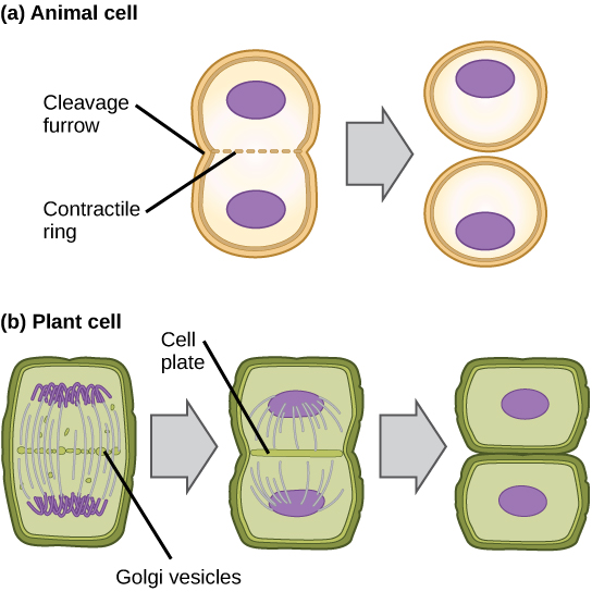
In cells such as animal cells that lack cell walls, cytokinesis begins following the onset of anaphase. A contractile ring composed of actin filaments forms just inside the plasma membrane at the former metaphase plate. The actin filaments pull the equator of the cell inward, forming a fissure. This fissure, or “crack,” is called the cleavage furrow    . The furrow deepens as the actin ring contracts, and eventually the membrane and cell are cleaved in two ( [link] ).

This illustration shows cytokinesis in a typical animal cell and a typical plant cell. In an animal cell, a contractile ring of actin filaments forms a cleavage furrow that divides the cell in two. In a plant cell, Golgi vesicles coalesce at the metaphase plate. A cell plate grows from the center outward, and the vesicles form a plasma membrane that divides the cytoplasm.
In part (a), a cleavage furrow forms at the former metaphase plate in the animal cell. The plasma membrane is drawn in by a ring of fibers contracting just inside the membrane. The cleavage furrow deepens until the cells are pinched in two.

Section summary

The cell cycle is an orderly sequence of events. Cells on the path to cell division proceed through a series of precisely timed and carefully regulated stages. In eukaryotes, the cell cycle consists of a long preparatory period, called interphase. Interphase is divided into G 1 , S, and G 2 phases. Mitosis consists of five stages: prophase, prometaphase, metaphase, anaphase, and telophase. Mitosis is usually accompanied by cytokinesis, during which the cytoplasmic components of the daughter cells are separated either by an actin ring (animal cells) or by cell plate formation (plant cells).

Art connections

[link] Which of the following is the correct order of events in mitosis?

  1. Sister chromatids line up at the metaphase plate. The kinetochore becomes attached to the mitotic spindle. The nucleus re-forms and the cell divides. The sister chromatids separate.
  2. The kinetochore becomes attached to the mitotic spindle. The sister chromatids separate. Sister chromatids line up at the metaphase plate. The nucleus re-forms and the cell divides.
  3. The kinetochore becomes attached to metaphase plate. Sister chromatids line up at the metaphase plate. The kinetochore breaks down and the sister chromatids separate. The nucleus re-forms and the cell divides.
  4. The kinetochore becomes attached to the mitotic spindle. Sister chromatids line up at the metaphase plate. The kinetochore breaks apart and the sister chromatids separate. The nucleus re-forms and the cell divides.

[link] D. The kinetochore becomes attached to the mitotic spindle. Sister chromatids line up at the metaphase plate. The kinetochore breaks apart and the sister chromatids separate. The nucleus reforms and the cell divides.

Got questions? Get instant answers now!

Questions & Answers

how does Neisseria cause meningitis
Nyibol Reply
what is microbiologist
Muhammad Reply
what is errata
Muhammad
is the branch of biology that deals with the study of microorganisms.
Ntefuni Reply
What is microbiology
Mercy Reply
studies of microbes
Louisiaste
when we takee the specimen which lumbar,spin,
Ziyad Reply
How bacteria create energy to survive?
Muhamad Reply
Bacteria doesn't produce energy they are dependent upon their substrate in case of lack of nutrients they are able to make spores which helps them to sustain in harsh environments
_Adnan
But not all bacteria make spores, l mean Eukaryotic cells have Mitochondria which acts as powerhouse for them, since bacteria don't have it, what is the substitution for it?
Muhamad
they make spores
Louisiaste
what is sporadic nd endemic, epidemic
Aminu Reply
the significance of food webs for disease transmission
Abreham
food webs brings about an infection as an individual depends on number of diseased foods or carriers dully.
Mark
explain assimilatory nitrate reduction
Esinniobiwa Reply
Assimilatory nitrate reduction is a process that occurs in some microorganisms, such as bacteria and archaea, in which nitrate (NO3-) is reduced to nitrite (NO2-), and then further reduced to ammonia (NH3).
Elkana
This process is called assimilatory nitrate reduction because the nitrogen that is produced is incorporated in the cells of microorganisms where it can be used in the synthesis of amino acids and other nitrogen products
Elkana
Examples of thermophilic organisms
Shu Reply
Give Examples of thermophilic organisms
Shu
advantages of normal Flora to the host
Micheal Reply
Prevent foreign microbes to the host
Abubakar
they provide healthier benefits to their hosts
ayesha
They are friends to host only when Host immune system is strong and become enemies when the host immune system is weakened . very bad relationship!
Mark
what is cell
faisal Reply
cell is the smallest unit of life
Fauziya
cell is the smallest unit of life
Akanni
ok
Innocent
cell is the structural and functional unit of life
Hasan
is the fundamental units of Life
Musa
what are emergency diseases
Micheal Reply
There are nothing like emergency disease but there are some common medical emergency which can occur simultaneously like Bleeding,heart attack,Breathing difficulties,severe pain heart stock.Hope you will get my point .Have a nice day ❣️
_Adnan
define infection ,prevention and control
Innocent
I think infection prevention and control is the avoidance of all things we do that gives out break of infections and promotion of health practices that promote life
Lubega
Heyy Lubega hussein where are u from?
_Adnan
en français
Adama
which site have a normal flora
ESTHER Reply
Many sites of the body have it Skin Nasal cavity Oral cavity Gastro intestinal tract
Safaa
skin
Asiina
skin,Oral,Nasal,GIt
Sadik
How can Commensal can Bacteria change into pathogen?
Sadik
How can Commensal Bacteria change into pathogen?
Sadik
all
Tesfaye
by fussion
Asiina
what are the advantages of normal Flora to the host
Micheal
what are the ways of control and prevention of nosocomial infection in the hospital
Micheal
what is inflammation
Shelly Reply
part of a tissue or an organ being wounded or bruised.
Wilfred
what term is used to name and classify microorganisms?
Micheal Reply
Binomial nomenclature
adeolu
Got questions? Join the online conversation and get instant answers!
Jobilize.com Reply

Get Jobilize Job Search Mobile App in your pocket Now!

Get it on Google Play Download on the App Store Now


Source:  OpenStax, Human biology. OpenStax CNX. Dec 01, 2015 Download for free at http://legacy.cnx.org/content/col11903/1.3
Google Play and the Google Play logo are trademarks of Google Inc.

Notification Switch

Would you like to follow the 'Human biology' conversation and receive update notifications?

Ask