

| << Chapter < Page | Chapter >> Page > |
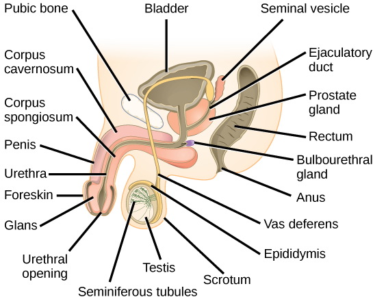
Sperm cell formation begins in the walls of seminiferous tubules that are coiled inside the testes ( [link] ; [link] ). The walls of the seminiferous tubules are made up of the developing sperm cells, with the least developed sperm at the periphery of the tubule; the cells get pushed closer to the lumen as maturation continues. The sperm cells are associated with Sertoli cells that nourish and promote the development of the sperm. Other cells present between the walls of the tubules are the Leydig/interstitial cells , which produce testosterone once the male reaches puberty.
When the sperm have developed flagella they leave the seminiferous tubules and enter the epididymis ( [link] ; [link] ). This structure lies along the top and back side of the testes and is the site of sperm maturation. The sperm leave the epididymis and enter the vas deferens, which carries the sperm behind the bladder, and forms the ejaculatory duct with the duct from the seminal vesicles. During a vasectomy, a section of the vas deferens is removed, preventing sperm (but not the secretions of the accessory glands) from being passed out of the body during ejaculation and preventing fertilization. Although a vasectomy is in many cases reversible via surgery, it is still considered to be a permanent procedure.
The bulk of the semen comes from the accessory glands associated with the male reproductive system. These are the seminal vesicles , the prostate gland , and the bulbourethral gland ( [link] ; [link] ). The secretions from the accessory glands provide important compounds for the sperm including nutrients, electrolytes, and pH buffering.
| Male Reproductive Anatomy | ||
|---|---|---|
| Organ | Location | Function |
| Scrotum | External | Supports testes and regulates their temperature |
| Penis | External | Delivers urine, copulating organ |
| Testes | Internal | Produce sperm and male hormones |
| Seminal Vesicles | Internal | Contribute to semen production |
| Prostate Gland | Internal | Contributes to semen production |
| Bulbourethtral Glands | Internal | Neutralize urine in urethra |
Gametogenesis, the production of sperm and eggs, involves the process of meiosis. During meiosis, two nuclear divisions separate the paired chromosomes in the nucleus and then separate the chromatids that were made during an earlier stage of the cell’s life cycle. Meiosis and its associated cell divisions produces haploid (n) cells with half of each pair of chromosomes normally found in diploid (2n)cells. The production of sperm is called spermatogenesis .
Spermatogenesis occurs in the wall of the seminiferous tubules, with the most primitive cells at the periphery of the tube and the most mature sperm at the lumen of the tube ( [link] ). Immediately under the capsule of the tubule are diploid, undifferentiated cells. These stem cells, each called a spermatogonium (pl. spermatogonia), go through mitosis to produce one cell that remains as a stem cell and a second cell called a primary spermatocyte that will undergo meiosis to produce sperm.
The diploid primary spermatocyte goes through meiosis I to produce two haploid cells called secondary spermatocytes. Each secondary spermatocyte divides after meiosis II to produce two cells called spermatids. The spermatids eventually reach the lumen of the tubule and grow a flagellum, becoming sperm cells. Four sperm result from each primary spermatocyte that goes through meiosis.
Notification Switch
Would you like to follow the 'Human biology' conversation and receive update notifications?

|
|

|
|

|
|
|
|
|

|
|

|
|

|
|

|
|

|
|

|