<< Chapter < Page Chapter >> Page >

Lysosomes

In animal cells, the lysosomes are the cell’s “garbage disposal.” Digestive enzymes within the lysosomes aid the breakdown of proteins, polysaccharides, lipids, nucleic acids, and even worn-out organelles. In single-celled eukaryotes, lysosomes are important for digestion of the food they ingest and the recycling of organelles. These enzymes are active at a much lower pH (more acidic) than those located in the cytoplasm. Many reactions that take place in the cytoplasm could not occur at a low pH, thus the advantage of compartmentalizing the eukaryotic cell into organelles is apparent.

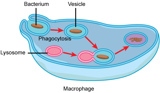
Lysosomes also use their hydrolytic enzymes to destroy disease-causing organisms that might enter the cell. A good example of this occurs in a group of white blood cells called macrophages, which are part of your body’s immune system. In a process known as phagocytosis, a section of the plasma membrane of the macrophage invaginates (folds in) and engulfs a pathogen. The invaginated section, with the pathogen inside, then pinches itself off from the plasma membrane and becomes a vesicle. The vesicle fuses with a lysosome. The lysosome’s hydrolytic enzymes then destroy the pathogen ( [link] ).

In this illustration, a eukaryotic cell is shown consuming a bacterium. As the bacterium is consumed, it is encapsulated into a vesicle. The vesicle fuses with a lysosome, and proteins inside the lysosome digest the bacterium.
A macrophage has phagocytized a potentially pathogenic bacterium into a vesicle, which then fuses with a lysosome within the cell so that the pathogen can be destroyed. Other organelles are present in the cell, but for simplicity, are not shown.

Vesicles

Vesicles are membrane-bound sacs that function in storage and transport. Vesicles can fuse with other membranes within the cell system.

Art connection

This figure shows the nucleus, rough ER, Golgi apparatus, vesicles, and plasma membrane. The right side of the rough ER is shown with an integral membrane protein embedded in it. The part of the protein facing the inside of the ER has a carbohydrate attached to it. The protein is shown leaving the ER in a vesicle that fuses with the cis face of the Golgi apparatus. The Golgi apparatus consists of several layers of membranes, called cisternae. As the protein passes through the cisternae, it is further modified by the addition of more carbohydrates. Eventually, it leaves the trans face of the Golgi in a vesicle. The vesicle fuses with the cell membrane so that the carbohydrate that was on the inside of the vesicle faces the outside of the membrane. At the same time, the contents of the vesicle are released from the cell.
The endomembrane system works to modify, package, and transport lipids and proteins. (credit: modification of work by Magnus Manske)

Ribosomes

Ribosomes are the cellular structures responsible for protein synthesis. When viewed through an electron microscope, free ribosomes appear as either clusters or single tiny dots floating freely in the cytoplasm. Ribosomes may be attached to either the cytoplasmic side of the plasma membrane or the cytoplasmic side of the endoplasmic reticulum. Electron microscopy has shown that ribosomes consist of large and small subunits. Ribosomes are enzyme complexes that are responsible for protein synthesis.

Because protein synthesis is essential for all cells, ribosomes are found in practically every cell, although they are smaller in prokaryotic cells. They are particularly abundant in immature red blood cells for the synthesis of hemoglobin, which functions in the transport of oxygen throughout the body.

Mitochondria

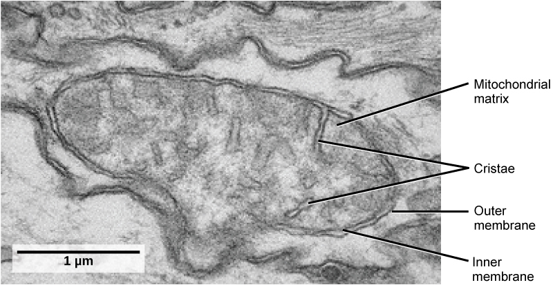
Mitochondria (singular = mitochondrion) are often called the “powerhouses” or “energy factories” of a cell because they are responsible for making adenosine triphosphate (ATP), the cell’s main energy-carrying molecule. The formation of ATP from the breakdown of glucose is known as cellular respiration. Mitochondria are oval-shaped, double-membrane organelles ( [link] ) that have their own ribosomes and DNA. Each membrane is a phospholipid bilayer embedded with proteins. The inner layer has folds called cristae, which increase the surface area of the inner membrane. The area surrounded by the folds is called the mitochondrial matrix. The cristae and the matrix have different roles in cellular respiration.

In keeping with our theme of form following function, it is important to point out that muscle cells have a very high concentration of mitochondria because muscle cells need a lot of energy to contract.

This transmission electron micrograph of a mitochondrion shows an oval, outer membrane and an inner membrane with many folds called cristae. Inside of the inner membrane is a space called the mitochondrial matrix.
This transmission electron micrograph shows a mitochondrion as viewed with an electron microscope. Notice the inner and outer membranes, the cristae, and the mitochondrial matrix. (credit: modification of work by Matthew Britton; scale-bar data from Matt Russell)

Section summary

Like a prokaryotic cell, a eukaryotic cell has a plasma membrane, cytoplasm, and ribosomes, but a eukaryotic cell is typically larger than a prokaryotic cell, has a true nucleus (meaning its DNA is surrounded by a membrane), and has other membrane-bound organelles that allow for compartmentalization of functions. The plasma membrane is a phospholipid bilayer embedded with proteins. The nucleolus within the nucleus is the site for ribosome assembly. Ribosomes are found in the cytoplasm or are attached to the cytoplasmic side of the plasma membrane or endoplasmic reticulum. They perform protein synthesis. Mitochondria perform cellular respiration and produce ATP. Vesicles are storage and transport compartments.

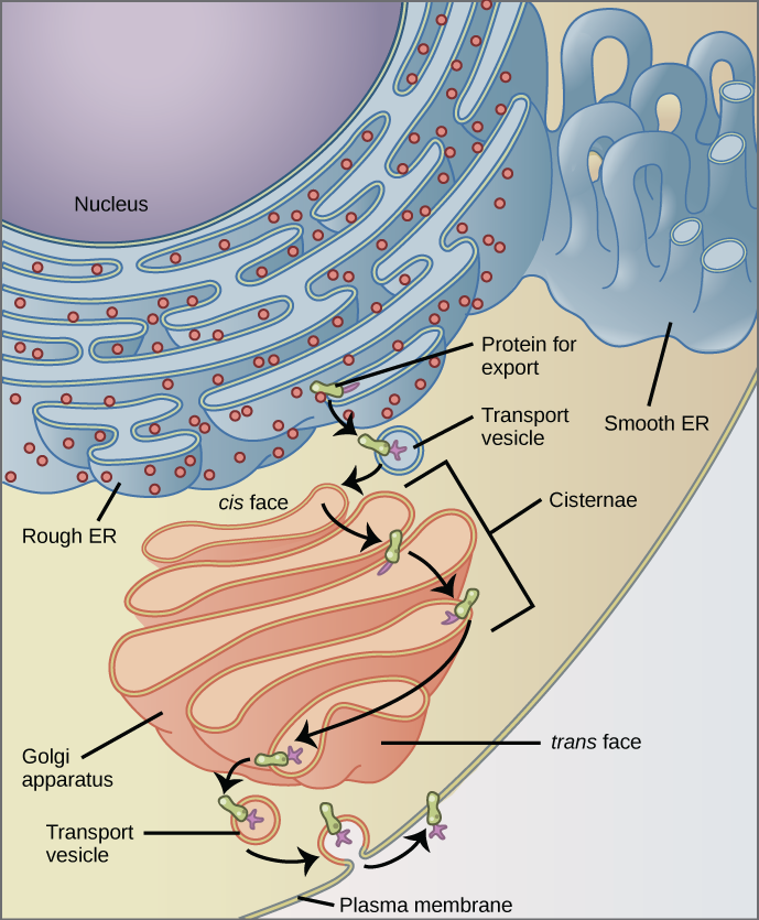
The endomembrane system includes the nuclear envelope, the endoplasmic reticulum, Golgi apparatus, lysosomes, vesicles, as well as the plasma membrane. These cellular components work together to modify, package, tag, and transport membrane lipids and proteins.

Art connections

[link] Why does the cis face of the Golgi not face the plasma membrane?

[link] Because that face receives chemicals from the ER, which is toward the center of the cell.

Got questions? Get instant answers now!

Questions & Answers

Ayele, K., 2003. Introductory Economics, 3rd ed., Addis Ababa.
Widad Reply
can you send the book attached ?
Ariel
?
Ariel
What is economics
Widad Reply
the study of how humans make choices under conditions of scarcity
AI-Robot
U(x,y) = (x×y)1/2 find mu of x for y
Desalegn Reply
U(x,y) = (x×y)1/2 find mu of x for y
Desalegn
what is ecnomics
Jan Reply
this is the study of how the society manages it's scarce resources
Belonwu
what is macroeconomic
John Reply
macroeconomic is the branch of economics which studies actions, scale, activities and behaviour of the aggregate economy as a whole.
husaini
etc
husaini
difference between firm and industry
husaini Reply
what's the difference between a firm and an industry
Abdul
firm is the unit which transform inputs to output where as industry contain combination of firms with similar production 😅😅
Abdulraufu
Suppose the demand function that a firm faces shifted from Qd  120 3P to Qd  90  3P and the supply function has shifted from QS  20  2P to QS 10  2P . a) Find the effect of this change on price and quantity. b) Which of the changes in demand and supply is higher?
Toofiq Reply
explain standard reason why economic is a science
innocent Reply
factors influencing supply
Petrus Reply
what is economic.
Milan Reply
scares means__________________ends resources. unlimited
Jan
economics is a science that studies human behaviour as a relationship b/w ends and scares means which have alternative uses
Jan
calculate the profit maximizing for demand and supply
Zarshad Reply
Why qualify 28 supplies
Milan
what are explicit costs
Nomsa Reply
out-of-pocket costs for a firm, for example, payments for wages and salaries, rent, or materials
AI-Robot
concepts of supply in microeconomics
David Reply
economic overview notes
Amahle Reply
identify a demand and a supply curve
Salome Reply
i don't know
Parul
there's a difference
Aryan
Demand curve shows that how supply and others conditions affect on demand of a particular thing and what percent demand increase whith increase of supply of goods
Israr
Hi Sir please how do u calculate Cross elastic demand and income elastic demand?
Abari
Got questions? Join the online conversation and get instant answers!
Jobilize.com Reply

Get Jobilize Job Search Mobile App in your pocket Now!

Get it on Google Play Download on the App Store Now


Source:  OpenStax, Human biology. OpenStax CNX. Dec 01, 2015 Download for free at http://legacy.cnx.org/content/col11903/1.3
Google Play and the Google Play logo are trademarks of Google Inc.

Notification Switch

Would you like to follow the 'Human biology' conversation and receive update notifications?

Ask