<< Chapter < Page Chapter >> Page >

Light microscopes commonly used in the undergraduate college laboratory magnify up to approximately 400 times. Two parameters that are important in microscopy are magnification and resolving power. Magnification is the process of enlarging an object in appearance. Resolving power is the ability of a microscope to distinguish two adjacent structures as separate: the higher the resolution, the better the clarity and detail of the image. When oil immersion lenses are used for the study of small objects, magnification is usually increased to 1,000 times. In order to gain a better understanding of cellular structure and function, scientists typically use electron microscopes.

Part a: This light microscope has binocular lenses and four objective lenses. The sample stage is directly beneath the objective lens. The light microscope sits on a tabletop and can be easily carried. Part b: The electron microscope shown here sits in a museum. It is about the size of a desk, and a person can sit in front of it to operate it. A column taller than a person rises from the center of the scope.
(a) Most light microscopes used in a college biology lab can magnify cells up to approximately 400 times and have a resolution of about 200 nanometers. (b) Electron microscopes provide a much higher magnification, 100,000x, and a have a resolution of 50 picometers. (credit a: modification of work by "GcG"/Wikimedia Commons; credit b: modification of work by Evan Bench)

Electron microscopes

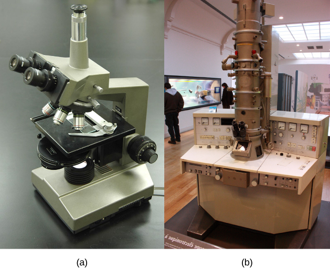
In contrast to light microscopes, electron microscopes ( [link] b ) use a beam of electrons instead of a beam of light. Not only does this allow for higher magnification and, thus, more detail ( [link] ), it also provides higher resolving power. The method used to prepare the specimen for viewing with an electron microscope kills the specimen. Electrons have short wavelengths (shorter than photons) that move best in a vacuum, so living cells cannot be viewed with an electron microscope.

In a scanning electron microscope, a beam of electrons moves back and forth across a cell’s surface, creating details of cell surface characteristics. In a transmission electron microscope, the electron beam penetrates the cell and provides details of a cell’s internal structures. As you might imagine, electron microscopes are significantly more bulky and expensive than light microscopes.

Part a: Salmonella through a light microscope appear as tiny purple dots. Part b: In this scanning electron micrograph, bacteria appear as three-dimensional ovals. The human cells are much larger with a complex, folded appearance. Some of the bacteria lie on the surface of the human cells, and some are squeezed between them.
(a) These Salmonella bacteria appear as tiny purple dots when viewed with a light microscope. (b) This scanning electron microscope micrograph shows Salmonella bacteria (in red) invading human cells (yellow). Even though subfigure (b) shows a different Salmonella specimen than subfigure (a), you can still observe the comparative increase in magnification and detail. (credit a: modification of work by CDC/Armed Forces Institute of Pathology, Charles N. Farmer, Rocky Mountain Laboratories; credit b: modification of work by NIAID, NIH; scale-bar data from Matt Russell)

For another perspective on cell size, try the HowBig interactive at this site .

Cell theory

The microscopes we use today are far more complex than those used in the 1600s by Antony van Leeuwenhoek, a Dutch shopkeeper who had great skill in crafting lenses. Despite the limitations of his now-ancient lenses, van Leeuwenhoek observed the movements of protista (a type of single-celled organism) and sperm, which he collectively termed “animalcules.”

In a 1665 publication called Micrographia , experimental scientist Robert Hooke coined the term “cell” for the box-like structures he observed when viewing cork tissue through a lens. In the 1670s, van Leeuwenhoek discovered bacteria and protozoa. Later advances in lenses, microscope construction, and staining techniques enabled other scientists to see some components inside cells.

By the late 1830s, botanist Matthias Schleiden and zoologist Theodor Schwann were studying tissues and proposed the unified cell theory    , which states that all living things are composed of one or more cells, the cell is the basic unit of life, and new cells arise from existing cells. Rudolf Virchow later made important contributions to this theory.

Career connection

Cytotechnologist

Have you ever heard of a medical test called a Pap smear ( [link] )? In this test, a doctor takes a small sample of cells from the uterine cervix of a patient and sends it to a medical lab where a cytotechnologist stains the cells and examines them for any changes that could indicate cervical cancer or a microbial infection.

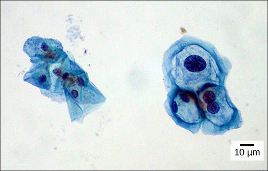
Cytotechnologists (cyto- = “cell”) are professionals who study cells via microscopic examinations and other laboratory tests. They are trained to determine which cellular changes are within normal limits and which are abnormal. Their focus is not limited to cervical cells; they study cellular specimens that come from all organs. When they notice abnormalities, they consult a pathologist, who is a medical doctor who can make a clinical diagnosis.

Cytotechnologists play a vital role in saving people’s lives. When abnormalities are discovered early, a patient’s treatment can begin sooner, which usually increases the chances of a successful outcome.

Both normal cells and cells infected with HPV have an irregular, round shape and a well-defined nucleus. Infected cells, however, are two to three times as large as uninfected cells, and some have two nuclei.
These uterine cervix cells, viewed through a light microscope, were obtained from a Pap smear. Normal cells are on the left. The cells on the right are infected with human papillomavirus (HPV). Notice that the infected cells are larger; also, two of these cells each have two nuclei instead of one, the normal number. (credit: modification of work by Ed Uthman, MD; scale-bar data from Matt Russell)

Section summary

A cell is the smallest unit of life. Most cells are so tiny that they cannot be seen with the naked eye. Therefore, scientists use microscopes to study cells. Electron microscopes provide higher magnification, higher resolution, and more detail than light microscopes. The unified cell theory states that all organisms are composed of one or more cells, the cell is the basic unit of life, and new cells arise from existing cells.

Questions & Answers

Ayele, K., 2003. Introductory Economics, 3rd ed., Addis Ababa.
Widad Reply
can you send the book attached ?
Ariel
?
Ariel
What is economics
Widad Reply
the study of how humans make choices under conditions of scarcity
AI-Robot
U(x,y) = (x×y)1/2 find mu of x for y
Desalegn Reply
U(x,y) = (x×y)1/2 find mu of x for y
Desalegn
what is ecnomics
Jan Reply
this is the study of how the society manages it's scarce resources
Belonwu
what is macroeconomic
John Reply
macroeconomic is the branch of economics which studies actions, scale, activities and behaviour of the aggregate economy as a whole.
husaini
etc
husaini
difference between firm and industry
husaini Reply
what's the difference between a firm and an industry
Abdul
firm is the unit which transform inputs to output where as industry contain combination of firms with similar production 😅😅
Abdulraufu
Suppose the demand function that a firm faces shifted from Qd  120 3P to Qd  90  3P and the supply function has shifted from QS  20  2P to QS 10  2P . a) Find the effect of this change on price and quantity. b) Which of the changes in demand and supply is higher?
Toofiq Reply
explain standard reason why economic is a science
innocent Reply
factors influencing supply
Petrus Reply
what is economic.
Milan Reply
scares means__________________ends resources. unlimited
Jan
economics is a science that studies human behaviour as a relationship b/w ends and scares means which have alternative uses
Jan
calculate the profit maximizing for demand and supply
Zarshad Reply
Why qualify 28 supplies
Milan
what are explicit costs
Nomsa Reply
out-of-pocket costs for a firm, for example, payments for wages and salaries, rent, or materials
AI-Robot
concepts of supply in microeconomics
David Reply
economic overview notes
Amahle Reply
identify a demand and a supply curve
Salome Reply
i don't know
Parul
there's a difference
Aryan
Demand curve shows that how supply and others conditions affect on demand of a particular thing and what percent demand increase whith increase of supply of goods
Israr
Hi Sir please how do u calculate Cross elastic demand and income elastic demand?
Abari
Got questions? Join the online conversation and get instant answers!
Jobilize.com Reply

Get Jobilize Job Search Mobile App in your pocket Now!

Get it on Google Play Download on the App Store Now


Source:  OpenStax, Biology. OpenStax CNX. Feb 29, 2016 Download for free at http://cnx.org/content/col11448/1.10
Google Play and the Google Play logo are trademarks of Google Inc.

Notification Switch

Would you like to follow the 'Biology' conversation and receive update notifications?

Ask