

| << Chapter < Page | Chapter >> Page > |
| Major Veins of the Brain | |
|---|---|
| Vessel | Description |
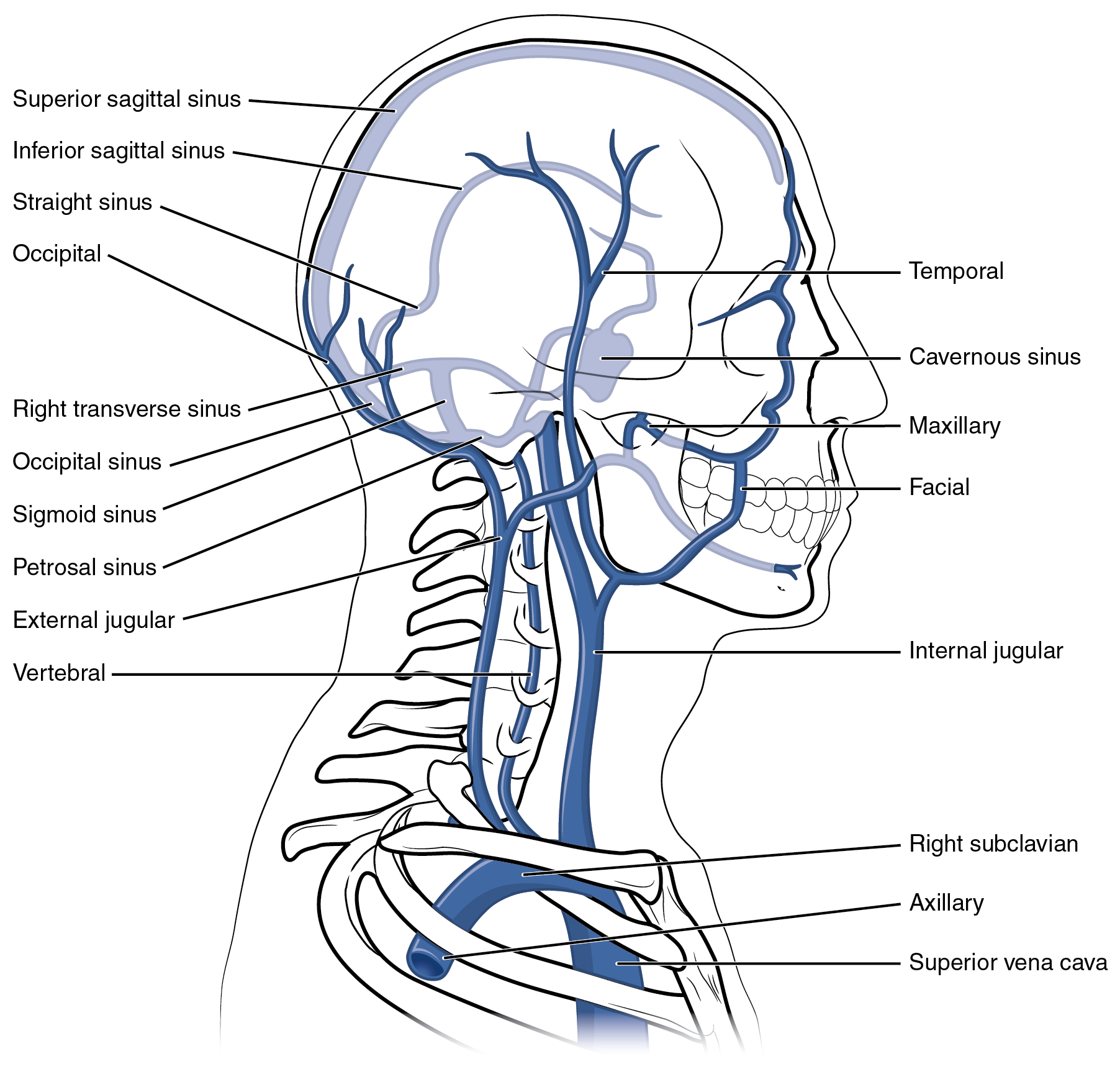
| Superior sagittal sinus | Enlarged vein located midsagittally between the meningeal and periosteal layers of the dura mater within the falx cerebri; receives most of the blood drained from the superior surface of the cerebrum and leads to the inferior jugular vein and the vertebral vein |
| Great cerebral vein | Receives most of the smaller vessels from the inferior cerebral veins and leads to the straight sinus |
| Straight sinus | Enlarged vein that drains blood from the brain; receives most of the blood from the great cerebral vein and leads to the left or right transverse sinus |
| Cavernous sinus | Enlarged vein that receives blood from most of the other cerebral veins and the eye socket, and leads to the petrosal sinus |
| Petrosal sinus | Enlarged vein that receives blood from the cavernous sinus and leads into the internal jugular veins |
| Occipital sinus | Enlarged vein that drains the occipital region near the falx cerebelli and leads to the left and right transverse sinuses, and also the vertebral veins |
| Transverse sinuses | Pair of enlarged veins near the lambdoid suture that drains the occipital, sagittal, and straight sinuses, and leads to the sigmoid sinuses |
| Sigmoid sinuses | Enlarged vein that receives blood from the transverse sinuses and leads through the jugular foramen to the internal jugular vein |
The digital veins in the fingers come together in the hand to form the palmar venous arches ( [link] ). From here, the veins come together to form the radial vein, the ulnar vein, and the median antebrachial vein. The radial vein and the ulnar vein parallel the bones of the forearm and join together at the antebrachium to form the brachial vein , a deep vein that flows into the axillary vein in the brachium.
The median antebrachial vein parallels the ulnar vein, is more medial in location, and joins the basilic vein in the forearm. As the basilic vein reaches the antecubital region, it gives off a branch called the median cubital vein that crosses at an angle to join the cephalic vein. The median cubital vein is the most common site for drawing venous blood in humans. The basilic vein continues through the arm medially and superficially to the axillary vein.
The cephalic vein begins in the antebrachium and drains blood from the superficial surface of the arm into the axillary vein. It is extremely superficial and easily seen along the surface of the biceps brachii muscle in individuals with good muscle tone and in those without excessive subcutaneous adipose tissue in the arms.
The subscapular vein drains blood from the subscapular region and joins the cephalic vein to form the axillary vein . As it passes through the body wall and enters the thorax, the axillary vein becomes the subclavian vein.
Many of the larger veins of the thoracic and abdominal region and upper limb are further represented in the flow chart in [link] . [link] summarizes the veins of the upper limbs.
Notification Switch
Would you like to follow the 'Anatomy & Physiology' conversation and receive update notifications?

|
|
|
|
|

|
|

|
|

|
|

|
|

|
|

|
|

|
|

|
|

|
|

|