

| << Chapter < Page | Chapter >> Page > |
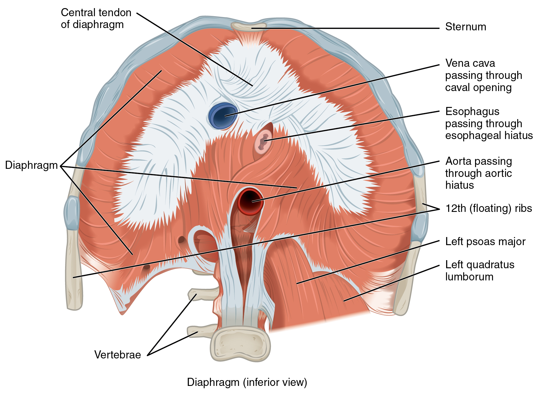
The change in volume of the thoracic cavity during breathing is due to the alternate contraction and relaxation of the diaphragm ( [link] ). It separates the thoracic and abdominal cavities, and is dome-shaped at rest. The superior surface of the diaphragm is convex, creating the elevated floor of the thoracic cavity. The inferior surface is concave, creating the curved roof of the abdominal cavity.
Defecating, urination, and even childbirth involve cooperation between the diaphragm and abdominal muscles (this cooperation is referred to as the “Valsalva maneuver”). You hold your breath by a steady contraction of the diaphragm; this stabilizes the volume and pressure of the peritoneal cavity. When the abdominal muscles contract, the pressure cannot push the diaphragm up, so it increases pressure on the intestinal tract (defecation), urinary tract (urination), or reproductive tract (childbirth).
The inferior surface of the pericardial sac and the inferior surfaces of the pleural membranes (parietal pleura) fuse onto the central tendon of the diaphragm. To the sides of the tendon are the skeletal muscle portions of the diaphragm, which insert into the tendon while having a number of origins including the xiphoid process of the sternum anteriorly, the inferior six ribs and their cartilages laterally, and the lumbar vertebrae and 12th ribs posteriorly.
The diaphragm also includes three openings for the passage of structures between the thorax and the abdomen. The inferior vena cava passes through the caval opening , and the esophagus and attached nerves pass through the esophageal hiatus. The aorta, thoracic duct, and azygous vein pass through the aortic hiatus of the posterior diaphragm.
There are three sets of muscles, called intercostal muscles , which span each of the intercostal spaces. The principal role of the intercostal muscles is to assist in breathing by changing the dimensions of the rib cage ( [link] ).
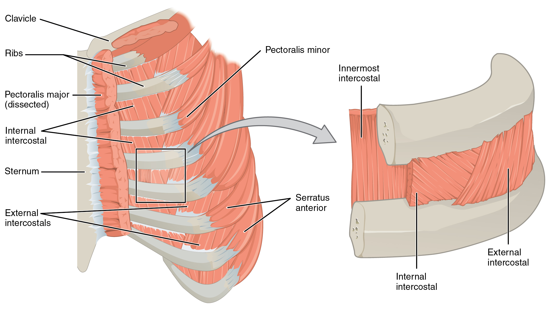
The 11 pairs of superficial external intercostal muscles aid in inspiration of air during breathing because when they contract, they raise the rib cage, which expands it. The 11 pairs of internal intercostal muscles, just under the externals, are used for expiration because they draw the ribs together to constrict the rib cage. The innermost intercostal muscles are the deepest, and they act as synergists for the action of the internal intercostals.
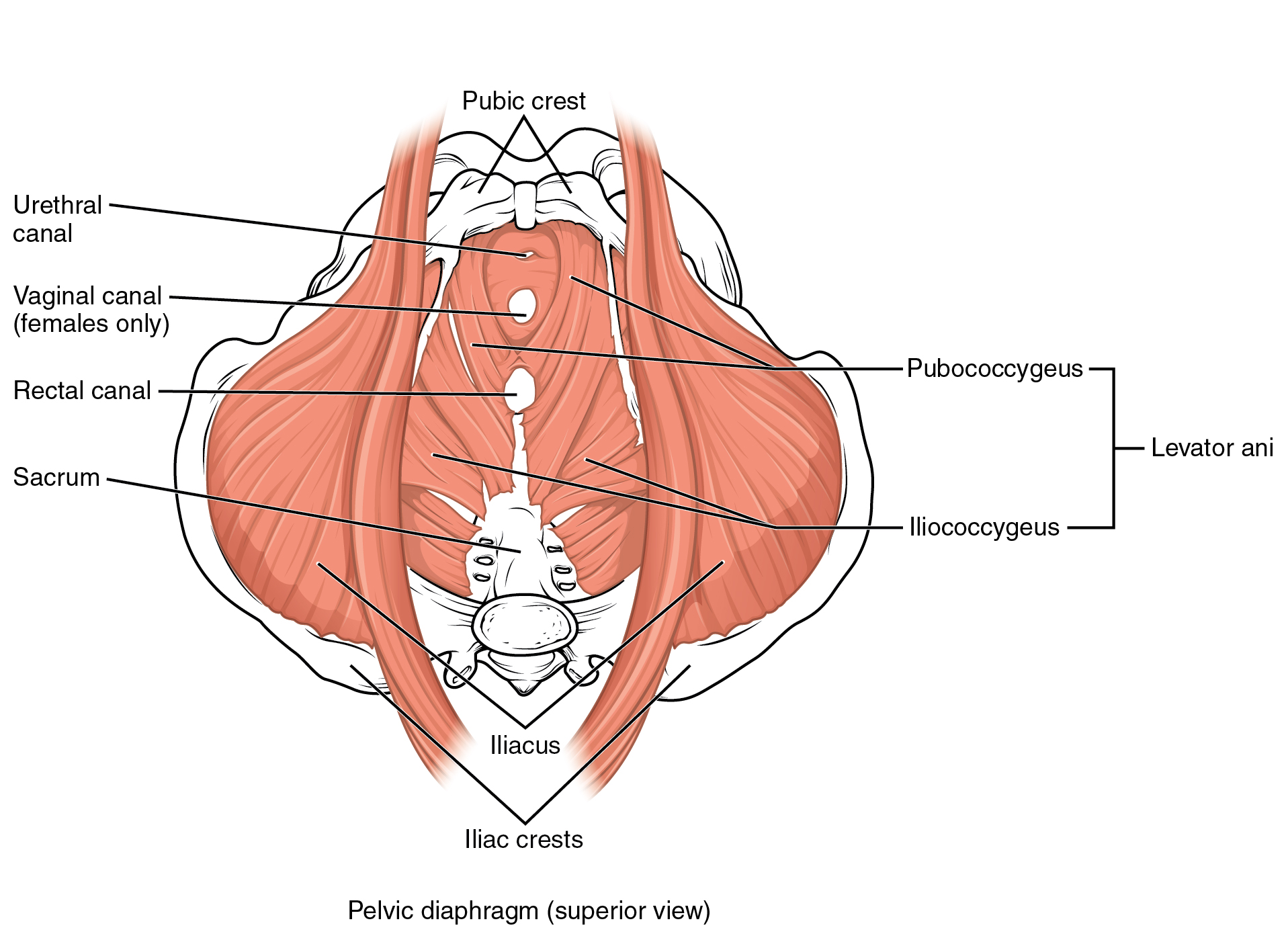
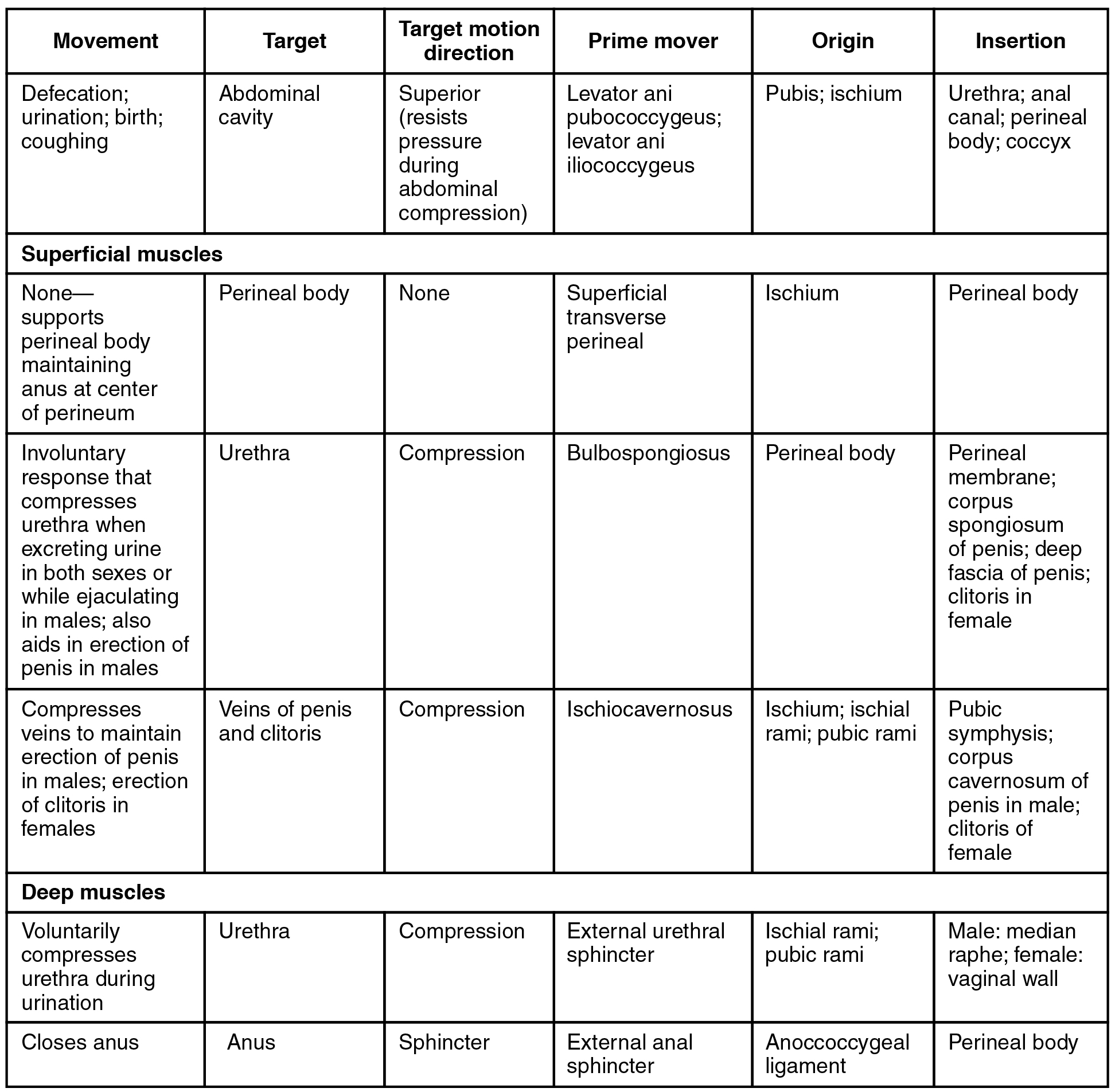
The pelvic floor is a muscular sheet that defines the inferior portion of the pelvic cavity. The pelvic diaphragm , spanning anteriorly to posteriorly from the pubis to the coccyx, comprises the levator ani and the ischiococcygeus. Its openings include the anal canal and urethra, and the vagina in women.
The large levator ani consists of two skeletal muscles, the pubococcygeus and the iliococcygeus ( [link] ). The levator ani is considered the most important muscle of the pelvic floor because it supports the pelvic viscera. It resists the pressure produced by contraction of the abdominal muscles so that the pressure is applied to the colon to aid in defecation and to the uterus to aid in childbirth (assisted by the ischiococcygeus , which pulls the coccyx anteriorly). This muscle also creates skeletal muscle sphincters at the urethra and anus.
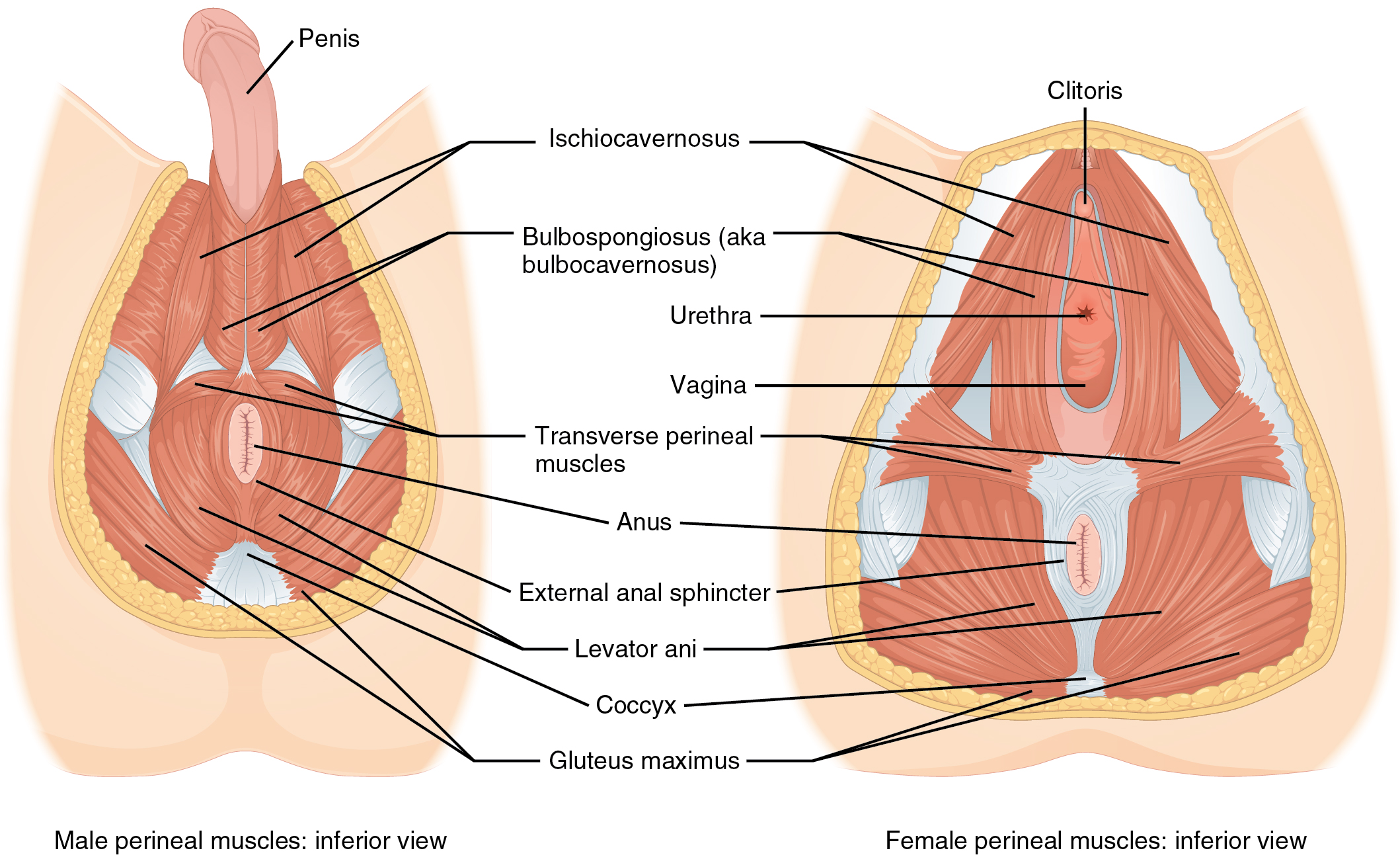
The perineum is the diamond-shaped space between the pubic symphysis (anteriorly), the coccyx (posteriorly), and the ischial tuberosities (laterally), lying just inferior to the pelvic diaphragm (levator ani and coccygeus). Divided transversely into triangles, the anterior is the urogenital triangle , which includes the external genitals. The posterior is the anal triangle , which contains the anus ( [link] ). The perineum is also divided into superficial and deep layers with some of the muscles common to men and women ( [link] ). Women also have the compressor urethrae and the sphincter urethrovaginalis , which function to close the vagina. In men, there is the deep transverse perineal muscle that plays a role in ejaculation.
Made of skin, fascia, and four pairs of muscle, the anterior abdominal wall protects the organs located in the abdomen and moves the vertebral column. These muscles include the rectus abdominis, which extends through the entire length of the trunk, the external oblique, the internal oblique, and the transversus abdominus. The quadratus lumborum forms the posterior abdominal wall.
The muscles of the thorax play a large role in breathing, especially the dome-shaped diaphragm. When it contracts and flattens, the volume inside the pleural cavities increases, which decreases the pressure within them. As a result, air will flow into the lungs. The external and internal intercostal muscles span the space between the ribs and help change the shape of the rib cage and the volume-pressure ratio inside the pleural cavities during inspiration and expiration.
The perineum muscles play roles in urination in both sexes, ejaculation in men, and vaginal contraction in women. The pelvic floor muscles support the pelvic organs, resist intra-abdominal pressure, and work as sphincters for the urethra, rectum, and vagina.
Notification Switch
Would you like to follow the 'Anatomy & Physiology' conversation and receive update notifications?

|
|

|
|

|
|

|
|

|
|

|
|

|
|

|
|

|
|

|
|
|
|
|
|