<< Chapter < Page Chapter >> Page >
By the end of this section, you will be able to:
  • Identify the components of the integumentary system
  • Describe the layers of the skin and the functions of each layer
  • Identify and describe the hypodermis and deep fascia
  • Describe the role of keratinocytes and their life cycle
  • Describe the role of melanocytes in skin pigmentation

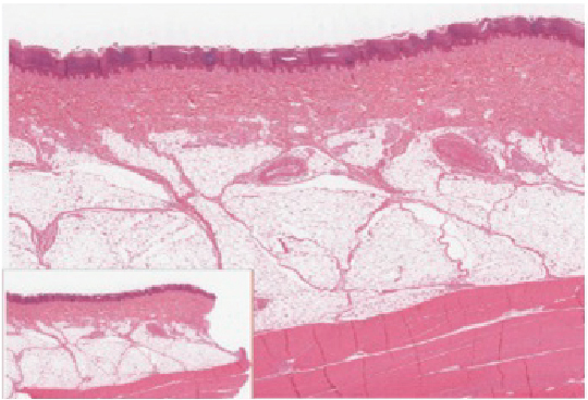
Although you may not typically think of the skin as an organ, it is in fact made of tissues that work together as a single structure to perform unique and critical functions. The skin and its accessory structures make up the integumentary system    , which provides the body with overall protection. The skin is made of multiple layers of cells and tissues, which are held to underlying structures by connective tissue ( [link] ). The deeper layer of skin is well vascularized (has numerous blood vessels). It also has numerous sensory, and autonomic and sympathetic nerve fibers ensuring communication to and from the brain.

Layers of skin

This illustration shows a cross section of skin tissue. The outermost layer is called the epidermis, and occupies one fifth of the cross section. Several hairs are emerging from the surface. The epidermis dives around one of the hairs, forming a follicle. The middle layer is called the dermis, which occupies four fifths of the cross section. The dermis contains an erector pilli muscle connected to one of the follicles. The dermis also contains an eccrine sweat gland, composed of a bunch of tubules. One tubule travels up from the bunch, through the epidermis, opening onto the surface a pore. There are two string-like nerves travelling vertically through the dermis. The right nerve is attached to a Pacinian corpuscle, which is a yellow structure consisting of concentric ovals similar to an onion. The lowest level of the skin, the hypodermis, contains fatty tissue, arteries, and veins. Blood vessels travel from the hypodermis and connect to hair follicles and erector pilli muscle in the dermis.
The skin is composed of two main layers: the epidermis, made of closely packed epithelial cells, and the dermis, made of dense, irregular connective tissue that houses blood vessels, hair follicles, sweat glands, and other structures. Beneath the dermis lies the hypodermis, which is composed mainly of loose connective and fatty tissues.

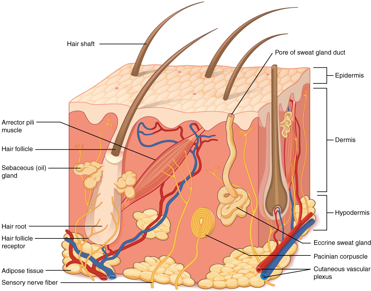
The skin consists of two main layers and a closely associated layer. View this animation to learn more about layers of the skin. What are the basic functions of each of these layers?

The epidermis

The epidermis    is composed of keratinized, stratified squamous epithelium. It is made of four or five layers of epithelial cells, depending on its location in the body. It does not have any blood vessels within it (i.e., it is avascular). Skin that has four layers of cells is referred to as “thin skin.” From deep to superficial, these layers are the stratum basale, stratum spinosum, stratum granulosum, and stratum corneum. Most of the skin can be classified as thin skin. “Thick skin” is found only on the palms of the hands and the soles of the feet. It has a fifth layer, called the stratum lucidum, located between the stratum corneum and the stratum granulosum ( [link] ).

Thin skin versus thick skin

Part A is a micrograph showing a cross section of thin skin. The topmost layer is a thin, translucent layer with irregular texture and areas where cells are sloughing off. The deepest layer is dark purple and extends into the third layer with finger like projections. The third light purple layer contains thin bands of fibers and small, dark cells. The fourth, and deepest layer, is darker than the third layer, but is still light purple. It contains thick fiber bands that are loosely packed. Part B is a magnified view of the epidermis of thick skin. It shows the topmost layer is five times thicker than the topmost layer of thin skin. The topmost layer of thick skin is also denser and less translucent than the topmost layer of thin skin.
These slides show cross-sections of the epidermis and dermis of (a) thin and (b) thick skin. Note the significant difference in the thickness of the epithelial layer of the thick skin. From top, LM × 40, LM × 40. (Micrographs provided by the Regents of University of Michigan Medical School © 2012)

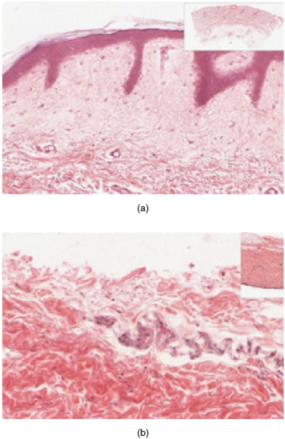
The cells in all of the layers except the stratum basale are called keratinocytes. A keratinocyte    is a cell that manufactures and stores the protein keratin. Keratin is an intracellular fibrous protein that gives hair, nails, and skin their hardness and water-resistant properties. The keratinocytes in the stratum corneum are dead and regularly slough away, being replaced by cells from the deeper layers ( [link] ).

Epidermis

The outer layer of cells in this micrograph is the thinnest layer and stained deep purple due to full keratinization of dead cells. The next layer occupies one quarter of the micrograph, is lightly stained, and is a dense collection of cells. The third layer from the top is mostly white, with lightly stained, loosely-packed strands radiating in random directions. The bottom-most layer is densely-packed, with thick bands of highly organized muscle tissue that are darkly stained.
The epidermis is epithelium composed of multiple layers of cells. The basal layer consists of cuboidal cells, whereas the outer layers are squamous, keratinized cells, so the whole epithelium is often described as being keratinized stratified squamous epithelium. LM × 40. (Micrograph provided by the Regents of University of Michigan Medical School © 2012)

Questions & Answers

calculate molarity of NaOH solution when 25.0ml of NaOH titrated with 27.2ml of 0.2m H2SO4
Gasin Reply
what's Thermochemistry
rhoda Reply
the study of the heat energy which is associated with chemical reactions
Kaddija
How was CH4 and o2 was able to produce (Co2)and (H2o
Edafe Reply
explain please
Victory
First twenty elements with their valences
Martine Reply
what is chemistry
asue Reply
what is atom
asue
what is the best way to define periodic table for jamb
Damilola Reply
what is the change of matter from one state to another
Elijah Reply
what is isolation of organic compounds
IKyernum Reply
what is atomic radius
ThankGod Reply
Read Chapter 6, section 5
Dr
Read Chapter 6, section 5
Kareem
Atomic radius is the radius of the atom and is also called the orbital radius
Kareem
atomic radius is the distance between the nucleus of an atom and its valence shell
Amos
Read Chapter 6, section 5
paulino
Bohr's model of the theory atom
Ayom Reply
is there a question?
Dr
when a gas is compressed why it becomes hot?
ATOMIC
It has no oxygen then
Goldyei
read the chapter on thermochemistry...the sections on "PV" work and the First Law of Thermodynamics should help..
Dr
Which element react with water
Mukthar Reply
Mgo
Ibeh
an increase in the pressure of a gas results in the decrease of its
Valentina Reply
definition of the periodic table
Cosmos Reply
What is the lkenes
Da Reply
what were atoms composed of?
Moses Reply
Got questions? Join the online conversation and get instant answers!
Jobilize.com Reply

Get Jobilize Job Search Mobile App in your pocket Now!

Get it on Google Play Download on the App Store Now


Source:  OpenStax, Anatomy & Physiology. OpenStax CNX. Feb 04, 2016 Download for free at http://legacy.cnx.org/content/col11496/1.8
Google Play and the Google Play logo are trademarks of Google Inc.

Notification Switch

Would you like to follow the 'Anatomy & Physiology' conversation and receive update notifications?

Ask