

| << Chapter < Page | Chapter >> Page > |
The intensity of the peak depends on the amount of material present, while the peak position is element dependent. Auger transitions characteristic of each elements can be found in the literature. Auger transitions of the first forty detectable elements are listed in [link] .
| Atomic number | Element | AES transition | Kinetic energy of transition (eV) |
|---|---|---|---|
| 3 | Li | KLL | 43 |
| 4 | Be | KLL | 104 |
| 5 | B | KLL | 179 |
| 6 | C | KLL | 272 |
| 7 | N | KLL | 379 |
| 8 | O | KLL | 508 |
| 9 | F | KLL | 647 |
| 11 | Na | KLL | 990 |
| 12 | Mg | KLL | 1186 |
| 13 | Al | LMM | 68 |
| 14 | Si | LMM | 92 |
| 15 | P | LMM | 120 |
| 16 | S | LMM | 152 |
| 17 | Cl | LMM | 181 |
| 19 | K | KLL | 252 |
| 20 | Ca | LMM | 291 |
| 21 | Sc | LMM | 340 |
| 22 | Ti | LMM | 418 |
| 23 | V | LMM | 473 |
| 24 | Cr | LMM | 529 |
| 25 | Mn | LMM | 589 |
| 26 | Fe | LMM | 703 |
| 27 | Co | LMM | 775 |
| 28 | Ni | LMM | 848 |
| 29 | Cu | LMM | 920 |
| 30 | Zn | LMM | 994 |
| 31 | Ga | LMM | 1070 |
| 32 | Ge | LMM | 1147 |
| 33 | As | LMM | 1228 |
| 34 | Se | LMM | 1315 |
| 35 | Br | LMM | 1376 |
| 39 | Y | MNN | 127 |
| 40 | Zr | MNN | 147 |
| 41 | Nb | MNN | 167 |
| 42 | Mo | MNN | 186 |
Important elements of an Auger spectrometer include a vacuum system, an electron source, and a detector. AES must be performed at pressures less than 10 -3 pascal (Pa) to keep residual gases from adsorbing to the sample surface. This can be achieved using an ultra-high-vacuum system with pressures from 10 -8 to 10 -9 Pa. Typical electron sources include tungsten filaments with an electron beam diameter of 3 - 5 μm, LaB 6 electron sources with a beam diameter of less than 40 nm, and Schottky barrier filaments with a 20 nm beam diameter and high beam current density. Two common detectors are the cylindrical mirror analyzer and the concentric hemispherical analyzer discussed below. Notably, concentric hemispherical analyzers typically have better energy resolution.
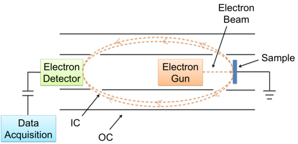
A CMA is composed of an electron gun, two cylinders, and an electron detector ( [link] ). The operation of a CMA involves an electron gun being directed at the sample. An ejected electron then enters the space between the inner and outer cylinders (IC and OC). The inner cylinder is at ground potential, while the outer cylinder’s potential is proportional to the kinetic energy of the electron. Due to its negative potential, the outer cylinder deflects the electron towards the electron detector. Only electrons within the solid angle cone are detected. The resulting signal is proportional to the number of electrons detected as a function of kinetic energy.
A CHA contains three parts ( [link] ):
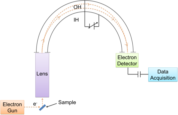
Electrons ejected from the surface enter the input lens, which focuses the electrons and retards their energy for better resolution. Electrons then enter the hemispheres through an entrance slit. A potential difference is applied on the hemispheres so that only electrons with a small range of energy differences reach the exit. Finally, an electron detector analyzes the electrons.
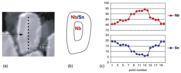
AES has widespread use owing to its ability to analyze small spot sizes with diameters from 5 μm down to 10 nm depending on the electron gun. For instance, AES is commonly employed to study film growth and surface-chemical composition, as well as grain boundaries in metals and ceramics. It is also used for quality control surface analyses in integrated circuit production lines due to short acquisition times. Moreover, AES is used for areas that require high spatial resolution, which XPS cannot achieve. AES can also be used in conjunction with transmission electron microscopy (TEM) and scanning electron microscopy (SEM) to obtain a comprehensive understanding of microscale materials, both chemically and structurally. As an example of combining techniques to investigate microscale materials, [link] shows the characterization of a single wire from a Sn-Nb multi-wire alloy. [link] a is a SEM image of the singular wire and [link] b is a schematic depicting the distribution of Nb and Sn within the wire. Point analysis was performed along the length of the wire to determine the percent concentrations of Nb and Sn.
AES is widely used for depth profiling. Depth profiling allows the elemental distributions of layered samples 0.2 – 1 μm thick to be characterized beyond the escape depth limit of an electron. Varying the incident and collection angles, and the primary beam energy controls the analysis depth. In general, the depth resolution decreases with the square root of the sample thickness. Notably, in AES, it is possible to simultaneously sputter and collect Auger data for depth profiling. The sputtering time indicates the depth and the intensity indicates elemental concentrations. Since, the sputtering process does not affect the ejection of the Auger electron, helium or argon ions can be used to sputter the surface and create the trench, while collecting Auger data at the same time. The depth profile does not have the problem of diffusion of hydrocarbons into the trenches. Thus, AES is better for depth profiles of reactive metals (e.g., gold or any metal or semiconductor). Yet, care should be taken because sputtering can mix up different elements, changing the sample composition.
While AES is a very valuable surface analysis technique, there are limitations. Because AES is a three-electron process, elements with less than three electrons cannot be analyzed. Therefore, hydrogen and helium cannot be detected. Nonetheless, detection is better for lighter elements with fewer transitions. The numerous transition peaks in heavier elements can cause peak overlap, as can the increased peak width of higher energy transitions. Detection limits of AES include 0.1 – 1% of a monolayer, 10 -16 – 10 -15 g of material, and 10 12 – 10 13 atoms/cm 2 .
Another limitation is sample destruction. Although focusing of the electron beam can improve resolution; the high-energy electrons can destroy the sample. To limit destruction, beam current densities of greater than 1 mA/cm 2 should be used. Furthermore, charging of the electron beam on insulating samples can deteriorate the sample and result in high-energy peak shifts or the appearance of large peaks.
Notification Switch
Would you like to follow the 'Physical methods in chemistry and nano science' conversation and receive update notifications?

|
|

|
|

|
|
|
|
|

|
|

|
|

|
|

|
|

|
|

|