<< Chapter < Page Chapter >> Page >

Though the signal’s position is well-defined according to the position of the xy mirrors, the signal from fluorescence is relatively weak after passing through the pinhole, so a photomultiplier tube is used to detect emitted photons. Detecting all photons without regard to spatial position increases the signal, and the photomultiplier tube further increases the detection signal by propagating an electron cascade resulting from the photoelectric effect (incident photons kicking off electrons). The resulting signal is an analog electrical signal with continuously varying voltage that corresponds to the emission intensity. This is periodically sampled by an analog-to-digital converter.

It is important to understand that the image is a reconstruction of many points sampled across the specimen. At any given time the microscope is only looking at a tiny point, and no complete image exists that can be viewed at an instantaneous point in time. Software is used to recombine these points to form an image plane, and combine image planes to form a 3-D representation of the sample volume.

Two-photon microscopy

Two-photon microscopy is a technique whereby two beams of lower intensity are directed to intersect at the focal point. Two photons can excite a fluorophore if they hit it at the same time, but alone they do not have enough energy to excite any molecules. The probability of two photons hitting a fluorophore at nearly the exact same time (less than 10 -16 ) is very low, but more likely at the focal point. This creates a bright point of light in the sample without the usual cone of light above and below the focal plane, since there are almost no excitations away from the focal point.

Schematic representation of the difference between single photon and two photon microscopy. Copyright: J. Mertz, Boston University.

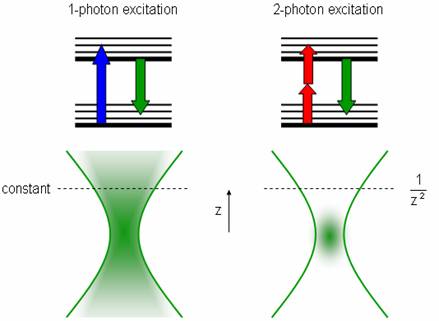
To increase the chance of absorption, an ultra-fast pulsed laser is used to create quick, intense light pulses. Since the hourglass shape is replaced by a point source, the pinhole near the detector (used to reduce the signal from light originating from outside the focal plane) can be eliminated. This also increases the signal-to-noise ratio (here is very little noise now that the light source is so focused, but the signal is also small). These lasers have lower average incident power than normal lasers, which helps reduce damage to the surrounding specimen. This technique can image deeper into the specimen (~400 μm), but these lasers are still very expensive, difficult to set up, require a stronger power supply, intensive cooling, and must be aligned in the same optical table because pulses can be distorted in optical fibers.

Microparticle characterization

Confocal microscopy is very useful for determining the relative positions of particles in three dimensions [link] . Software allows measurement of distances in the 3D reconstructions so that information about spacing can be ascertained (such as packing density, porosity, long range order or alignment, etc.).

A reconstruction of a colloidal suspension of poly(methyl methacrylate) (PMMA) microparticles approximately 2 microns in diameter. Adapted from Confocal Microscopy of Colloids , Eric Weeks.

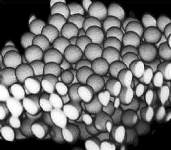
If imaging in fluorescence mode, remember that the signal will only represent the locations of the individual fluorophores. There is no guarantee fluorophores will completely attach to the structures of interest or that there will not be stray fluorophores away from those structures. For microparticles it is often possible to attach the fluorophores to the shell of the particle, creating hollow spheres of fluorophores. It is possible to tell if a sample sphere is hollow or solid but it would depend on the transparency of the material.

Dispersions of microparticles have been used to study nucleation and crystal growth, since colloids are much larger than atoms and can be imaged in real-time. Crystalline regions are determined from the order of spheres arranged in a lattice, and regions can be distinguished from one another by noting lattice defects.

Self-assembly is another application where time-dependent, 3-D studies can help elucidate the assembly process and determine the position of various structures or materials. Because confocal is popular for biological specimens, the position of nanoparticles such as quantum dots in a cell or tissue can be observed. This can be useful for determining toxicity, drug-delivery effectiveness, diffusion limitations, etc.

A summary of confocal microscopy’s strengths and weaknesses

    Strengths

  • Less haze, better contrast than ordinary microscopes.
  • 3-D capability.
  • Illuminates a small volume.
  • Excludes most of the light from the sample not in the focal plane.
  • Depth of field may be adjusted with pinhole size.
  • Has both reflected light and fluorescence modes.
  • Can image living cells and tissues.
  • Fluorescence microscopy can identify several different structures simultaneously.
  • Accommodates samples with thickness up to 100 μm.
  • Can use with two-photon microscopy.
  • Allows for optical sectioning (no artifacts from physical sectioning) 0.5 - 1.5 μm.

    Weakness

  • Images are scanned slowly (one complete image every 0.1-1 second).
  • Must raster scan sample, no complete image exists at any given time.
  • There is an inherent resolution limit because of diffraction (based on numerical aperture, ~200 nm).
  • Sample should be relatively transparent for good signal.
  • High fluorescence concentrations can quench the fluorescent signal.
  • Fluorophores irreversibly photobleach.
  • Lasers are expensive.
  • Angle of incident light changes slightly, introducing slight distortion.

Bibliography

  • P. Davidovits and M. D. Egger, Nature , 1973, 244 , 366.
  • A. Hibbs, Confocal Microscopy for Biologists , Twayne Publishers, Boston (2004).
  • M. Minsky, Scanning , 1988, 10 , 128.
  • J. Pawley, Handbook of Biological Confocal Microscopy , Twayne Publishers, Boston (2006).
  • V. Prasad, D. Semwogerere, and E.R. Weeks, J. Phys.: Condens. Matter , 2007, 19 , 113102.
  • D. Semwogerere and E. R. Weeks, Encyclopedia of Biomaterials and Biomedical Engineering Confocal Microscopy , Taylor Francis (2005).
  • C. Sheppard, Confocal Laser Scanning Microscopy , Twayne Publishers, Boston (1997).

Questions & Answers

summarize halerambos & holbon
David Reply
the Three stages of Auguste Comte
Clementina Reply
what are agents of socialization
Antonio Reply
sociology of education
Nuhu Reply
definition of sociology of education
Nuhu
what is culture
Abdulrahim Reply
shared beliefs, values, and practices
AI-Robot
What are the two type of scientific method
ogunniran Reply
I'm willing to join you
Aceng Reply
what are the scientific method of sociology
Man
what is socialization
ogunniran Reply
the process wherein people come to understand societal norms and expectations, to accept society's beliefs, and to be aware of societal values
AI-Robot
scientific method in doing research
ogunniran
defimition of sickness in afica
Anita
Cosmology
ogunniran
Hmmm
ogunniran
list and explain the terms that found in society
REMMY Reply
list and explain the terms that found in society
Mukhtar
what are the agents of socialization
Antonio
Family Peer group Institution
Abdulwajud
I mean the definition
Antonio
ways of perceived deviance indifferent society
Naomi Reply
reasons of joining groups
SAM
to bring development to the nation at large
Hyellafiya
entails of consultative and consensus building from others
Gadama
World first Sociologist?
Abu
What is evolutionary model
Muhammad Reply
Evolution models refer to mathematical and computational representations of the processes involved in biological evolution. These models aim to simulate and understand how species change over time through mechanisms such as natural selection, genetic drift, and mutation. Evolutionary models can be u
faruk
what are the modern trends in religious behaviours
Selekeye Reply
what are social norms
Daniel Reply
shared standards of acceptable behavior by the group or appropriate behavior in a particular institution or those behaviors that are acceptable in a society
Lucius
that is how i understood it
Lucius
examples of societal norms
Diamond
Discuss the characteristics of the research located within positivist and the interpretivist paradigm
Tariro Reply
what is Industrialisation
Selekeye Reply
industrialization
Angelo
Got questions? Join the online conversation and get instant answers!
Jobilize.com Reply

Get Jobilize Job Search Mobile App in your pocket Now!

Get it on Google Play Download on the App Store Now


Source:  OpenStax, Physical methods in chemistry and nano science. OpenStax CNX. May 05, 2015 Download for free at http://legacy.cnx.org/content/col10699/1.21
Google Play and the Google Play logo are trademarks of Google Inc.

Notification Switch

Would you like to follow the 'Physical methods in chemistry and nano science' conversation and receive update notifications?

Ask