<< Chapter < Page Chapter >> Page >
The graph shows the variation of intensity as a function of sine theta. The curve has a strong peak at sine theta equals zero, then has small oscillations spreading symmetrically to the left and right of this central peak. The oscillations all appear to be of the same height. Between each oscillation, the curve appears to go to zero, and each zero is labeled. The first zero to the left of the main peak is labeled minus lambda over d and the first zero to the right is labeled lambda over d. The second zero to the left is labeled minus two lambda over d and the second zero to the right is labeled two lambda over d. The third zero to the left is labeled minus three lambda over d and the third zero to the right is labeled three lambda over d.
A graph of single slit diffraction intensity showing the central maximum to be wider and much more intense than those to the sides. In fact the central maximum is six times higher than shown here.

Thus, to obtain destructive interference for a single slit    ,
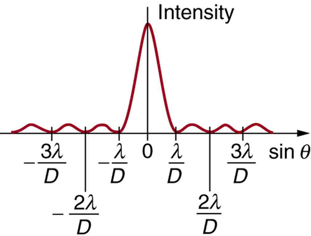
D sin θ = , for m = 1, –1, 2, –2, 3, (destructive), size 12{D`"sin"θ= ital "mλ",~m="1,"`"2,"`"3,"` dotslow } {}

where D size 12{D} {} is the slit width, λ size 12{λ} {} is the light’s wavelength, θ size 12{θ} {} is the angle relative to the original direction of the light, and m size 12{m} {} is the order of the minimum. [link] shows a graph of intensity for single slit interference, and it is apparent that the maxima on either side of the central maximum are much less intense and not as wide. This is consistent with the illustration in [link] (b).

Calculating single slit diffraction

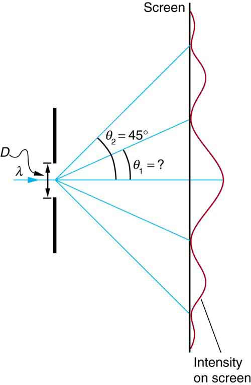
Visible light of wavelength 550 nm falls on a single slit and produces its second diffraction minimum at an angle of 45.0º size 12{"45" "." 0°} {} relative to the incident direction of the light. (a) What is the width of the slit? (b) At what angle is the first minimum produced?

The schematic shows a single slit to the left and the resulting intensity pattern on a screen is graphed on the right. The single slit is represented by a gap of size d in a vertical line. A ray of wavelength lambda enters the gap from the left, then five rays leave from the gap center and head to the right. One ray continues on the horizontal centerline of the schematic. Two rays angle upward: the first at an unknown angle theta one above the horizontal and the second at an angle theta two equals forty five degrees above the horizontal. The final two rays angle downward at the same angles, so that they are symmetric about the horizontal with respect to the two rays that angle upward. The intensity on the screen is a maximum where the central ray hits the screen, whereas it is a minimum where the angled rays hit the screen.
A graph of the single slit diffraction pattern is analyzed in this example.

Strategy

From the given information, and assuming the screen is far away from the slit, we can use the equation D sin θ = size 12{D`"sin"θ= ital "mλ"} {} first to find D size 12{D} {} , and again to find the angle for the first minimum θ 1 size 12{θ rSub { size 8{1} } } {} .

Solution for (a)

We are given that λ = 550 nm size 12{λ="500"`"nm"} {} , m = 2 size 12{m=2} {} , and θ 2 = 45.0º . Solving the equation D sin θ = size 12{D`"sin"θ= ital "mλ"} {} for D size 12{D} {} and substituting known values gives

D = sin θ 2 = 2 ( 550 nm ) sin 45.0º = 1100 × 10 9 0.707 = 1.56 × 10 6 .

Solution for (b)

Solving the equation D sin θ = size 12{D`"sin"θ= ital "mλ"} {} for sin θ 1 size 12{"sin"θ rSub { size 8{1} } } {} and substituting the known values gives

sin θ 1 = D = 1 550 × 10 9 m 1 . 56 × 10 6 m . size 12{"sin"θ rSub { size 8{1} } = { {mλ} over {D} } = { {1 left ("550" times "10" rSup { size 8{ - 9} } m right )} over {1 "." "56" times "10" rSup { size 8{ - 6} } m} } } {}

Thus the angle θ 1 size 12{θ rSub { size 8{1} } } {} is

θ 1 = sin 1 0.354 = 20.7º. size 12{θ rSub { size 8{1} } ="sin" rSup { size 8{ - 1} } 0 "." "354"="20" "." 7°} {}

Discussion

We see that the slit is narrow (it is only a few times greater than the wavelength of light). This is consistent with the fact that light must interact with an object comparable in size to its wavelength in order to exhibit significant wave effects such as this single slit diffraction pattern. We also see that the central maximum extends 20.7º on either side of the original beam, for a width of about 41º . The angle between the first and second minima is only about 24º ( 45.0º 20.7º ) size 12{"24"°` \( "45" "." 0° - "20" "." 7° \) } {} . Thus the second maximum is only about half as wide as the central maximum.

Got questions? Get instant answers now!

Section summary

  • A single slit produces an interference pattern characterized by a broad central maximum with narrower and dimmer maxima to the sides.
  • There is destructive interference for a single slit when D sin θ = , (for m = 1, –1, 2, –2, 3, …) size 12{D`"sin"θ= ital "mλ",~m="1,"`"2,"`"3,"` dotslow } {} , where D is the slit width, λ is the light’s wavelength, θ is the angle relative to the original direction of the light, and m is the order of the minimum. Note that there is no m = 0 size 12{m=0} {} minimum.

Conceptual questions

As the width of the slit producing a single-slit diffraction pattern is reduced, how will the diffraction pattern produced change?

Got questions? Get instant answers now!

Problems&Exercises

(a) At what angle is the first minimum for 550-nm light falling on a single slit of width 1 . 00 μm size 12{1 "." "00"`"μm"} {} ? (b) Will there be a second minimum?

(a) 33 . size 12{"33" "." 4°} {}

(b) No

Got questions? Get instant answers now!

(a) Calculate the angle at which a 2 . 00 -μm size 12{2 "." "00""-μm"} {} -wide slit produces its first minimum for 410-nm violet light. (b) Where is the first minimum for 700-nm red light?

Got questions? Get instant answers now!

Questions & Answers

what is biology
Hajah Reply
the study of living organisms and their interactions with one another and their environments
AI-Robot
what is biology
Victoria Reply
HOW CAN MAN ORGAN FUNCTION
Alfred Reply
the diagram of the digestive system
Assiatu Reply
allimentary cannel
Ogenrwot
How does twins formed
William Reply
They formed in two ways first when one sperm and one egg are splited by mitosis or two sperm and two eggs join together
Oluwatobi
what is genetics
Josephine Reply
Genetics is the study of heredity
Misack
how does twins formed?
Misack
What is manual
Hassan Reply
discuss biological phenomenon and provide pieces of evidence to show that it was responsible for the formation of eukaryotic organelles
Joseph Reply
what is biology
Yousuf Reply
the study of living organisms and their interactions with one another and their environment.
Wine
discuss the biological phenomenon and provide pieces of evidence to show that it was responsible for the formation of eukaryotic organelles in an essay form
Joseph Reply
what is the blood cells
Shaker Reply
list any five characteristics of the blood cells
Shaker
lack electricity and its more savely than electronic microscope because its naturally by using of light
Abdullahi Reply
advantage of electronic microscope is easily and clearly while disadvantage is dangerous because its electronic. advantage of light microscope is savely and naturally by sun while disadvantage is not easily,means its not sharp and not clear
Abdullahi
cell theory state that every organisms composed of one or more cell,cell is the basic unit of life
Abdullahi
is like gone fail us
DENG
cells is the basic structure and functions of all living things
Ramadan
What is classification
ISCONT Reply
is organisms that are similar into groups called tara
Yamosa
in what situation (s) would be the use of a scanning electron microscope be ideal and why?
Kenna Reply
A scanning electron microscope (SEM) is ideal for situations requiring high-resolution imaging of surfaces. It is commonly used in materials science, biology, and geology to examine the topography and composition of samples at a nanoscale level. SEM is particularly useful for studying fine details,
Hilary
cell is the building block of life.
Condoleezza Reply
Got questions? Join the online conversation and get instant answers!
Jobilize.com Reply
Practice Key Terms 1

Get Jobilize Job Search Mobile App in your pocket Now!

Get it on Google Play Download on the App Store Now


Source:  OpenStax, College physics. OpenStax CNX. Jul 27, 2015 Download for free at http://legacy.cnx.org/content/col11406/1.9
Google Play and the Google Play logo are trademarks of Google Inc.

Notification Switch

Would you like to follow the 'College physics' conversation and receive update notifications?

Ask