<< Chapter < Page Chapter >> Page >
  • Discuss the single slit diffraction pattern.

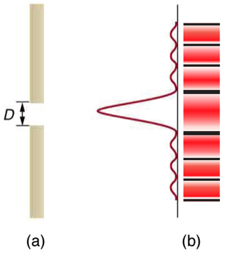
Light passing through a single slit forms a diffraction pattern somewhat different from those formed by double slits or diffraction gratings. [link] shows a single slit diffraction pattern. Note that the central maximum is larger than those on either side, and that the intensity decreases rapidly on either side. In contrast, a diffraction grating produces evenly spaced lines that dim slowly on either side of center.

Part a of the figure shows a slit in a vertical bar. To the right of the bar is a graph of intensity versus height. The graph is turned ninety degrees counterclockwise so that the intensity scale increases to the left and the height increases as you go up the page. Just in front of the gap, a strong central peak extends leftward from the graph’s baseline, and many smaller satellite peaks appear above and below this central peak. Part b of the figure shows a drawing of the two-dimensional intensity pattern that is observed from single slit diffraction. The central stripe is quite broad compared to the satellite stripes, and there are dark areas between all the stripes.
(a) Single slit diffraction pattern. Monochromatic light passing through a single slit has a central maximum and many smaller and dimmer maxima on either side. The central maximum is six times higher than shown. (b) The drawing shows the bright central maximum and dimmer and thinner maxima on either side.

The analysis of single slit diffraction is illustrated in [link] . Here we consider light coming from different parts of the same slit. According to Huygens’s principle, every part of the wavefront in the slit emits wavelets. These are like rays that start out in phase and head in all directions. (Each ray is perpendicular to the wavefront of a wavelet.) Assuming the screen is very far away compared with the size of the slit, rays heading toward a common destination are nearly parallel. When they travel straight ahead, as in [link] (a), they remain in phase, and a central maximum is obtained. However, when rays travel at an angle θ size 12{θ} {} relative to the original direction of the beam, each travels a different distance to a common location, and they can arrive in or out of phase. In [link] (b), the ray from the bottom travels a distance of one wavelength λ size 12{λ} {} farther than the ray from the top. Thus a ray from the center travels a distance λ / 2 size 12{λ/2} {} farther than the one on the left, arrives out of phase, and interferes destructively. A ray from slightly above the center and one from slightly above the bottom will also cancel one another. In fact, each ray from the slit will have another to interfere destructively, and a minimum in intensity will occur at this angle. There will be another minimum at the same angle to the right of the incident direction of the light.

The figure shows four schematics of a ray bundle passing through a single slit. The slit is represented as a gap in a vertical line. In the first schematic, the ray bundle passes horizontally through the slit. This schematic is labeled theta equals zero and bright. The second schematic is labeled dark and shows the ray bundle passing through the slit an angle of roughly fifteen degrees above the horizontal. The path length difference between the top and bottom ray is lambda, and the schematic is labeled sine theta equals lambda over d. The third schematic is labeled bright and shows the ray bundle passing through the slit at an angle of about twenty five degrees above the horizontal. The path length difference between the top and bottom rays is three lambda over two d, and the schematic is labeled sine theta equals three lambda over two d. The final schematic is labeled dark and shows the ray bundle passing through the slit at an angle of about forty degrees above the horizontal. The path length difference between the top and bottom rays is two lambda over d, and the schematic is labeled sine theta equals two lambda over d.
Light passing through a single slit is diffracted in all directions and may interfere constructively or destructively, depending on the angle. The difference in path length for rays from either side of the slit is seen to be D sin θ size 12{D`"sin"`θ} {} .

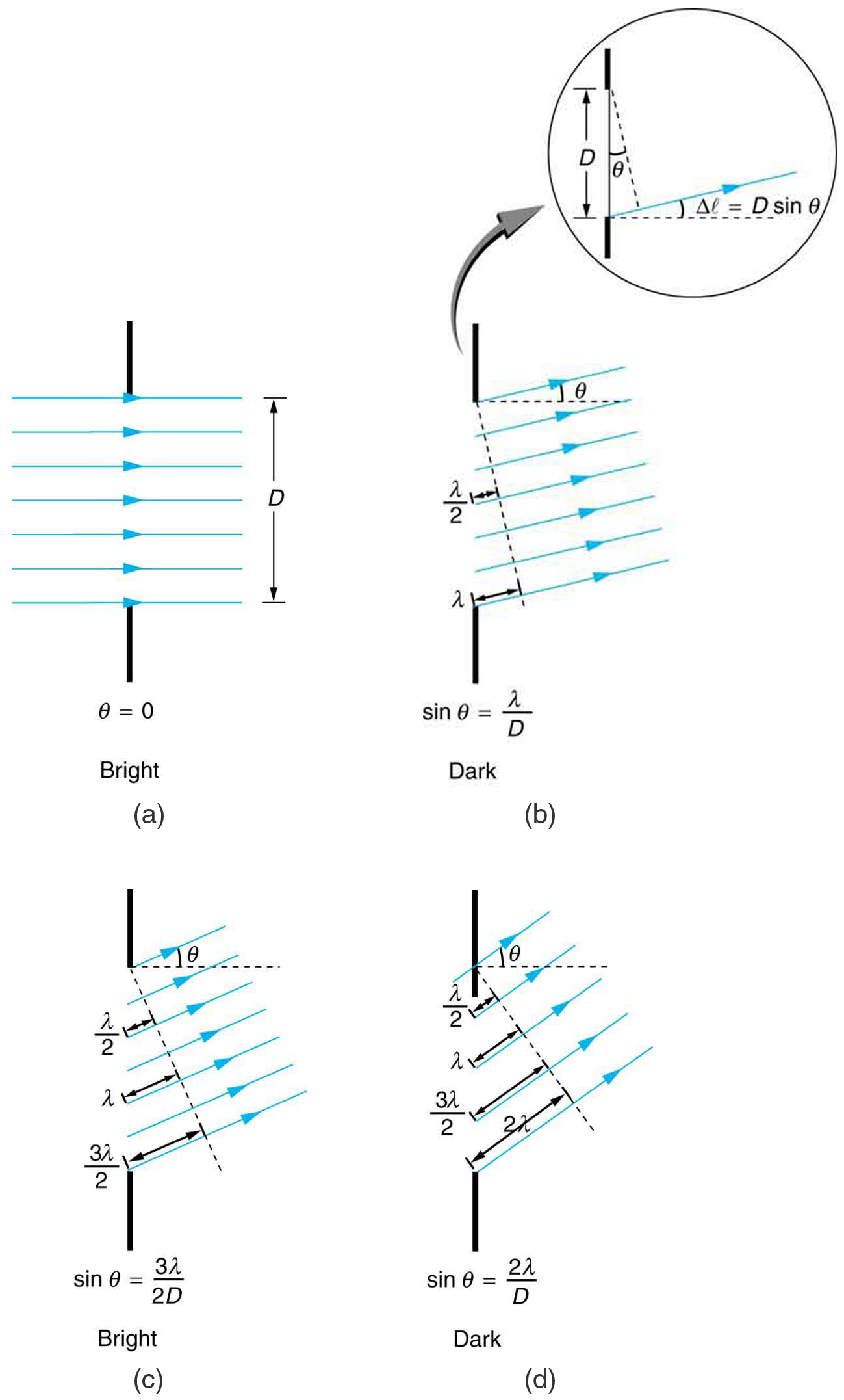
At the larger angle shown in [link] (c), the path lengths differ by / 2 size 12{3λ/2} {} for rays from the top and bottom of the slit. One ray travels a distance λ size 12{λ} {} different from the ray from the bottom and arrives in phase, interfering constructively. Two rays, each from slightly above those two, will also add constructively. Most rays from the slit will have another to interfere with constructively, and a maximum in intensity will occur at this angle. However, all rays do not interfere constructively for this situation, and so the maximum is not as intense as the central maximum. Finally, in [link] (d), the angle shown is large enough to produce a second minimum. As seen in the figure, the difference in path length for rays from either side of the slit is D sin θ size 12{D`"sin"θ} {} , and we see that a destructive minimum is obtained when this distance is an integral multiple of the wavelength.

Questions & Answers

what is biology
Hajah Reply
the study of living organisms and their interactions with one another and their environments
AI-Robot
what is biology
Victoria Reply
HOW CAN MAN ORGAN FUNCTION
Alfred Reply
the diagram of the digestive system
Assiatu Reply
allimentary cannel
Ogenrwot
How does twins formed
William Reply
They formed in two ways first when one sperm and one egg are splited by mitosis or two sperm and two eggs join together
Oluwatobi
what is genetics
Josephine Reply
Genetics is the study of heredity
Misack
how does twins formed?
Misack
What is manual
Hassan Reply
discuss biological phenomenon and provide pieces of evidence to show that it was responsible for the formation of eukaryotic organelles
Joseph Reply
what is biology
Yousuf Reply
the study of living organisms and their interactions with one another and their environment.
Wine
discuss the biological phenomenon and provide pieces of evidence to show that it was responsible for the formation of eukaryotic organelles in an essay form
Joseph Reply
what is the blood cells
Shaker Reply
list any five characteristics of the blood cells
Shaker
lack electricity and its more savely than electronic microscope because its naturally by using of light
Abdullahi Reply
advantage of electronic microscope is easily and clearly while disadvantage is dangerous because its electronic. advantage of light microscope is savely and naturally by sun while disadvantage is not easily,means its not sharp and not clear
Abdullahi
cell theory state that every organisms composed of one or more cell,cell is the basic unit of life
Abdullahi
is like gone fail us
DENG
cells is the basic structure and functions of all living things
Ramadan
What is classification
ISCONT Reply
is organisms that are similar into groups called tara
Yamosa
in what situation (s) would be the use of a scanning electron microscope be ideal and why?
Kenna Reply
A scanning electron microscope (SEM) is ideal for situations requiring high-resolution imaging of surfaces. It is commonly used in materials science, biology, and geology to examine the topography and composition of samples at a nanoscale level. SEM is particularly useful for studying fine details,
Hilary
cell is the building block of life.
Condoleezza Reply
Got questions? Join the online conversation and get instant answers!
Jobilize.com Reply
Practice Key Terms 1

Get Jobilize Job Search Mobile App in your pocket Now!

Get it on Google Play Download on the App Store Now


Source:  OpenStax, College physics. OpenStax CNX. Jul 27, 2015 Download for free at http://legacy.cnx.org/content/col11406/1.9
Google Play and the Google Play logo are trademarks of Google Inc.

Notification Switch

Would you like to follow the 'College physics' conversation and receive update notifications?

Ask