

| << Chapter < Page | Chapter >> Page > |
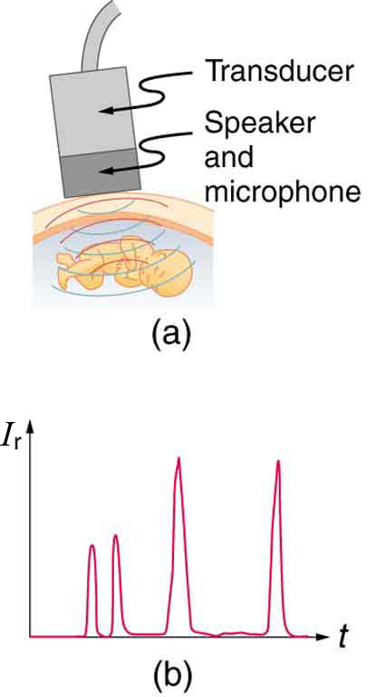
where and are the acoustic impedances of the two media making up the boundary. A reflection coefficient of zero (corresponding to total transmission and no reflection) occurs when the acoustic impedances of the two media are the same. An impedance “match” (no reflection) provides an efficient coupling of sound energy from one medium to another. The image formed in an ultrasound is made by tracking reflections (as shown in [link] ) and mapping the intensity of the reflected sound waves in a two-dimensional plane.
(a) Using the values for density and the speed of ultrasound given in [link] , show that the acoustic impedance of fat tissue is indeed .
(b) Calculate the intensity reflection coefficient of ultrasound when going from fat to muscle tissue.
Strategy for (a)
The acoustic impedance can be calculated using and the values for and found in [link] .
Solution for (a)
(1) Substitute known values from [link] into .
(2) Calculate to find the acoustic impedance of fat tissue.
This value is the same as the value given for the acoustic impedance of fat tissue.
Strategy for (b)
The intensity reflection coefficient for any boundary between two media is given by , and the acoustic impedance of muscle is given in [link] .
Solution for (b)
Substitute known values into to find the intensity reflection coefficient:
Discussion
This result means that only 1.4% of the incident intensity is reflected, with the remaining being transmitted.
The applications of ultrasound in medical diagnostics have produced untold benefits with no known risks. Diagnostic intensities are too low (about ) to cause thermal damage. More significantly, ultrasound has been in use for several decades and detailed follow-up studies do not show evidence of ill effects, quite unlike the case for x-rays.
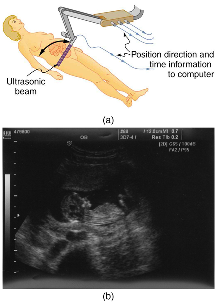
The most common ultrasound applications produce an image like that shown in [link] . The speaker-microphone broadcasts a directional beam, sweeping the beam across the area of interest. This is accomplished by having multiple ultrasound sources in the probe’s head, which are phased to interfere constructively in a given, adjustable direction. Echoes are measured as a function of position as well as depth. A computer constructs an image that reveals the shape and density of internal structures.
Notification Switch
Would you like to follow the 'College physics' conversation and receive update notifications?

|
|
|
|
|

|
|

|
|

|
|

|
|

|
|

|
|

|
|

|
|

|
|

|