<< Chapter < Page Chapter >> Page >
By the end of this section, you will be able to:
  • Identify the locations and primary secretions involved in the chemical digestion of carbohydrates, proteins, lipids, and nucleic acids
  • Compare and contrast absorption of the hydrophilic and hydrophobic nutrients

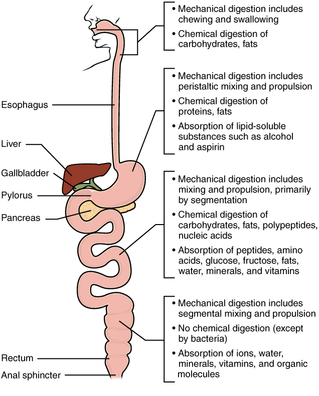
As you have learned, the process of mechanical digestion    is relatively simple. It involves the physical breakdown of food but does not alter its chemical makeup. Chemical digestion , on the other hand, is a complex process that reduces food into its chemical building blocks, which are then absorbed to nourish the cells of the body ( [link] ). In this section, you will look more closely at the processes of chemical digestion and absorption.

Digestion and absorption

This diagram identifies the functions of mechanical and chemical digestion and absorption at each organ. Next to each organ, a callout identifies which steps of digestion take place in that particular organ.
Digestion begins in the mouth and continues as food travels through the small intestine. Most absorption occurs in the small intestine.

Chemical digestion

Large food molecules (for example, proteins, lipids, nucleic acids, and starches) must be broken down into subunits that are small enough to be absorbed by the lining of the alimentary canal. This is accomplished by enzymes.

Carbohydrate digestion

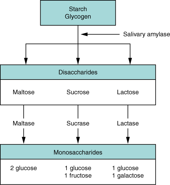
The average American diet is about 50 percent carbohydrates, which may be classified according to the number of monomers (subunits) they contain. You should take notes on figure 2 below .

The chemical digestion of starches begins in the mouth and has been reviewed in previous modules. In the small intestine, pancreatic amylase    does the ‘heavy lifting’ for starch and carbohydrate digestion ( [link] ). Amylases break down starches into simple sugars. Three brush border enzymes break up the sugars sucrose, lactose, and maltose into monosaccharides.

Carbohydrate digestion flow chart

This flow chart shows the steps in digestion of carbohydrates. The different levels shown are starch and glycogen, disaccharides and monosaccharides. Under each type of sugar, examples and the enzymes responsible for digestion are listed.
Carbohydrates are broken down into their monomers in a series of steps.

Protein digestion

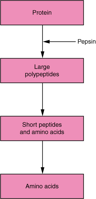
Proteins are polymers composed of amino acids linked by peptide bonds to form long chains. Digestion reduces them to their constituent amino acids. You usually consume about 15 to 20 percent of your total calorie intake as protein.

The digestion of protein starts in the stomach, where HCl and pepsin break proteins into smaller subunits, which then travel to the small intestine ( [link] ). Chemical digestion in the small intestine is continued by pancreatic enzymes, including chymotrypsin and trypsin , each of which act on specific bonds in amino acid sequences. At the same time, the cells of the brush border secrete enzymes. This results in molecules small enough to enter the bloodstream ( [link] ). Take notes on figure 4 .

Digestion of protein

This diagrams shows the human digestive system and identifies the role of each organ in protein digestion. A text call-out next to each organ details the specific function.
The digestion of protein begins in the stomach and is completed in the small intestine.

Digestion of protein flow chart

This flow chart shows the different steps in the digestion of protein. The four steps shown are protein, large polypeptides, short peptides and amino acids and amino acids.
Proteins are successively broken down into their amino acid components.

Lipid digestion

A healthy diet limits lipid intake to 35 percent of total calorie intake. The most common dietary lipids are triglycerides, which are made up of a glycerol molecule bound to three fatty acid chains. Small amounts of dietary cholesterol and phospholipids are also consumed.

The lipase primarily responsible for lipid digestion is pancreatic lipase    . However, because the pancreas is the only consequential source of lipase, virtually all lipid digestion occurs in the small intestine. Pancreatic lipase breaks down each triglyceride into subunits called free fatty acids and monoglycerides .

Questions & Answers

what is phylogeny
Odigie Reply
evolutionary history and relationship of an organism or group of organisms
AI-Robot
ok
Deng
what is biology
Hajah Reply
the study of living organisms and their interactions with one another and their environments
AI-Robot
what is biology
Victoria Reply
HOW CAN MAN ORGAN FUNCTION
Alfred Reply
the diagram of the digestive system
Assiatu Reply
allimentary cannel
Ogenrwot
How does twins formed
William Reply
They formed in two ways first when one sperm and one egg are splited by mitosis or two sperm and two eggs join together
Oluwatobi
what is genetics
Josephine Reply
Genetics is the study of heredity
Misack
how does twins formed?
Misack
What is manual
Hassan Reply
discuss biological phenomenon and provide pieces of evidence to show that it was responsible for the formation of eukaryotic organelles
Joseph Reply
what is biology
Yousuf Reply
the study of living organisms and their interactions with one another and their environment.
Wine
discuss the biological phenomenon and provide pieces of evidence to show that it was responsible for the formation of eukaryotic organelles in an essay form
Joseph Reply
what is the blood cells
Shaker Reply
list any five characteristics of the blood cells
Shaker
lack electricity and its more savely than electronic microscope because its naturally by using of light
Abdullahi Reply
advantage of electronic microscope is easily and clearly while disadvantage is dangerous because its electronic. advantage of light microscope is savely and naturally by sun while disadvantage is not easily,means its not sharp and not clear
Abdullahi
cell theory state that every organisms composed of one or more cell,cell is the basic unit of life
Abdullahi
is like gone fail us
DENG
cells is the basic structure and functions of all living things
Ramadan
What is classification
ISCONT Reply
is organisms that are similar into groups called tara
Yamosa
in what situation (s) would be the use of a scanning electron microscope be ideal and why?
Kenna Reply
A scanning electron microscope (SEM) is ideal for situations requiring high-resolution imaging of surfaces. It is commonly used in materials science, biology, and geology to examine the topography and composition of samples at a nanoscale level. SEM is particularly useful for studying fine details,
Hilary
Got questions? Join the online conversation and get instant answers!
Jobilize.com Reply

Get Jobilize Job Search Mobile App in your pocket Now!

Get it on Google Play Download on the App Store Now


Source:  OpenStax, Digestive system. OpenStax CNX. Feb 23, 2015 Download for free at http://legacy.cnx.org/content/col11761/1.1
Google Play and the Google Play logo are trademarks of Google Inc.

Notification Switch

Would you like to follow the 'Digestive system' conversation and receive update notifications?

Ask