<< Chapter < Page Chapter >> Page >
By the end of this section, you will be able to:
  • Explain the function of cartilage
  • List the steps of intramembranous ossification
  • List the steps of endochondral ossification
  • Explain the growth activity at the epiphyseal plate
  • Compare and contrast the processes of modeling and remodeling

In the early stages of embryonic development, the embryo’s skeleton consists of membranes hyaline cartilage. By the sixth or seventh week of embryonic life, the actual process of bone development, ossification    , begins. There are two ways that bones undergo ossification— intramembranous ossification    and endochondral ossification    —but bone is the same regardless of the pathway that produces it.

Cartilage templates

Throughout fetal development and into childhood growth and development, bone forms on the cartilaginous matrix. By the time a fetus is born, most of the cartilage has been replaced with bone. Some additional cartilage will be replaced throughout childhood, and some cartilage remains in the adult skeleton.

Intramembranous ossification

During intramembranous ossification    , compact and spongy bone develops directly from sheets of connective tissue. The flat bones of the face, most of the cranial bones, and the clavicles (collarbones) are formed via intramembranous ossification.

The process begins when cells in the embryonic skeleton gather together and begin to differentiate into specialized cells ( [link] a ). Some of these cells will differentiate into capillaries, while others will become osteogenic cells and then osteoblasts. Although they will ultimately be spread out by the formation of bone tissue, early osteoblasts appear in a cluster called an ossification center    .

The osteoblasts secrete osteoid    , uncalcified matrix, which calcifies (hardens) within a few days as mineral salts are deposited on it, thereby entrapping the osteoblasts within. Once entrapped, the osteoblasts become osteocytes ( [link] b ). As osteoblasts transform into osteocytes, osteogenic cells in the surrounding connective tissue differentiate into new osteoblasts.

Intramembranous ossification

Image A shows seven osteoblasts, cells with small, finger like projections. They are surrounded by granules of osteoid. Both the cells and the osteoid are contained within a blue, circular, ossification center that is surrounded by a “socket” of dark, string-like collagen fibers and gray mesenchymal cells. The cells are generally amorphous, similar in appearance to an amoeba. In image B, the ossification center is no longer surrounded by a ring of osteoblasts. The osteoblasts have secreted bone into the ossification center, creating a new bone matrix. There are also five osteocytes embedded in the new bone matrix. The osteocytes are thin, oval-shaped cells with many fingerlike projections. Osteoid particles are still embedded in the bony matrix in image B.  In image C, the ring of osteoblasts surrounding the ossification center has separated, forming an upper and lower layer of osteoblasts sandwiched between the two layers of mesenchyme cells. A label indicates that the mesenchyme cells and the surrounding collagen fibers form the periosteum. The osteoblasts secrete spongy bone into the space between the two osteoblast rows. Therefore, the accumulating spongy bone pushes the upper and lower rows of osteoblasts away from each other. In this image, most of the spongy bone has been secreted by the osteoblasts, as the trabeculae are visible. In addition, an artery has already broken through the periosteum and invaded the spongy bone. Image D looks similar to image C, except that the rows of osteoblasts are now secreting layers of compact bone between the spongy bone and the periosteum. The artery has now branched and spread throughout the spongy bone. A label indicates that the cavities between the trabeculae now contain red bone marrow.
Intramembranous ossification follows four steps. (a) Mesenchymal cells group into clusters, and ossification centers form. (b) Secreted osteoid traps osteoblasts, which then become osteocytes. (c) Trabecular matrix and periosteum form. (d) Compact bone develops superficial to the trabecular bone, and crowded blood vessels condense into red marrow.

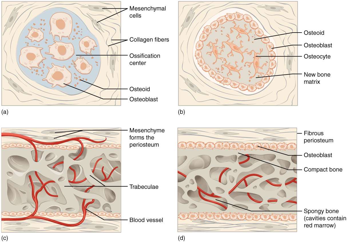
Intramembranous ossification begins during fetal development and continues on into adolescence. At birth, the skull and clavicles are not fully ossified nor are the sutures of the skull closed. This allows the skull and shoulders to deform during passage through the birth canal. The last bones to ossify via intramembranous ossification are the flat bones of the face, which reach their adult size at the end of the adolescent growth spurt.

Endochondral ossification

In endochondral ossification    , bone develops by replacing hyaline cartilage. Cartilage does not become bone. Instead, cartilage serves as a template or blueprint to be completely replaced by new bone. Endochondral ossification takes much longer than intramembranous ossification. Bones at the base of the skull and long bones form via endochondral ossification.

Get Jobilize Job Search Mobile App in your pocket Now!

Get it on Google Play Download on the App Store Now


Source:  OpenStax, Skeletal system. OpenStax CNX. Apr 17, 2015 Download for free at https://legacy.cnx.org/content/col11779/1.1
Google Play and the Google Play logo are trademarks of Google Inc.

Notification Switch

Would you like to follow the 'Skeletal system' conversation and receive update notifications?

Ask