<< Chapter < Page Chapter >> Page >
By the end of this section, you will be able to:
  • Explain the structure and function of epithelial tissue
  • Distinguish between tight junctions, anchoring junctions, and gap junctions
  • Distinguish between simple epithelia and stratified epithelia, as well as between squamous, cuboidal, and columnar epithelia
  • Describe the structure and function of endocrine and exocrine glands and their respective secretions

Most epithelial tissues are essentially large sheets of cells covering all the surfaces of the body exposed to the outside world and lining the outside of organs. Epithelium also forms much of the glandular tissue of the body. Skin is not the only area of the body exposed to the outside. Other areas include the airways, the digestive tract, as well as the urinary and reproductive systems, all of which are lined by an epithelium. Hollow organs and body cavities that do not connect to the exterior of the body, which includes, blood vessels and serous membranes, are also lined with epithelium.

All epithelia share some important structural and functional features. This tissue is highly cellular, meaning there are many cells with very little material between them. Adjoining cells form a connection between their cell membranes called a cell junction    . A basement membrane    is found at the bottom of the epithelia and helps to hold the cells in place.

Generalized functions of epithelial tissue

Epithelial tissues provide the body’s first line of protection from physical, chemical, and biological wear and tear. The cells of an epithelium act as gatekeepers of the body controlling permeability allowing only certain substances to pass through it. All substances that enter the body must cross an epithelium.

Many epithelial cells are capable of secretion and release mucous and other compounds. The epithelium of the small intestine releases digestive enzymes, for example. Cells lining the respiratory tract secrete mucous that traps incoming microorganisms and particles.

    The functions of epithelia

  • Protects underlying structures. Skin protects the delicate tissues beneath it from scrapes.
  • Acts as a barrier. Skin keeps bacteria out of your body.
  • Permits passage of substances. The air sacs of the lung allow oxygen and carbon dioxide to pass through them.
  • Secretes substances. Epithelia that line the nasal passages produce mucus.
  • Absorbs substances. The epithelia in the intestines absorb nutrients from the food into the blood.

Cell to cell junctions

Types of cell junctions

These three illustrations each show the edges of two vertical cell membranes. The cell membranes are viewed partially from the side so that the inside edge of the right cell membrane is visible. The upper left image shows a tight junction. The two cell membranes are bound by transmembrane protein strands. The proteins travel the inside edge of the right cell membrane and cross over to the left cell membrane, cinching the two membranes together. The cell membranes are still somewhat separated in between neighboring strands, creating intercellular spaces. The upper right diagram shows a gap junction. The gap junctions are composed of two interlocking connexins, which are round, hollow tubes that extend through the cell membranes. Two connexins, one from the left cell membrane and the other from the right cell membrane, meet between the two cells, forming a connexon. Even at the site of the connexon, there is a small gap between the cell membranes. On the inside edge of the right cell membrane, the gap junction appears as a depression. Three connexins are embedded into the membranes like buttons on a shirt. The bottom images show the three types of anchoring junctions. The left image shows a desmosome. Here, the inside edge of both the right and left cell membranes have brown, round plaques. Each plaque has tentacle-like intermediate filaments (keratin) that extend into each cell’s cytoplasm. The two plaques are connected across the intercellular space by several interlocking transmembrane glycoproteins (cadherin). The connected glycoproteins look similar to a zipped-up zipper between the right and left cell membranes. The right image shows an adheren. These are similar to desmosomes, with two plaques on the inside edge of each cell membrane connected across the intercellular space by glycoproteins. However, the plaques do not contain the tentacle-like intermediate filaments branching into the cytoplasm. Instead, the plaques are ribbed with green actin filaments. The filaments are neatly arranged in parallel, horizontal strands on the surface of the plaque facing the cytoplasm. The bottom image shows a hemidesmosome. Rather than located between two neighboring cells, the hemidesmosome is located between the bottom of a cell and the basement membrane. A hemidesmosome contains a single plaque on the inside edge of the cell membrane. Like the desmosome, intermediate filaments project from the plaque into the cytoplasm. The opposite side of the plaque has purple, knob-shaped integrins extending out to the basal lamina of the basement membrane.
The three basic types of cell-to-cell junctions are tight junctions, gap junctions, and anchoring junctions.

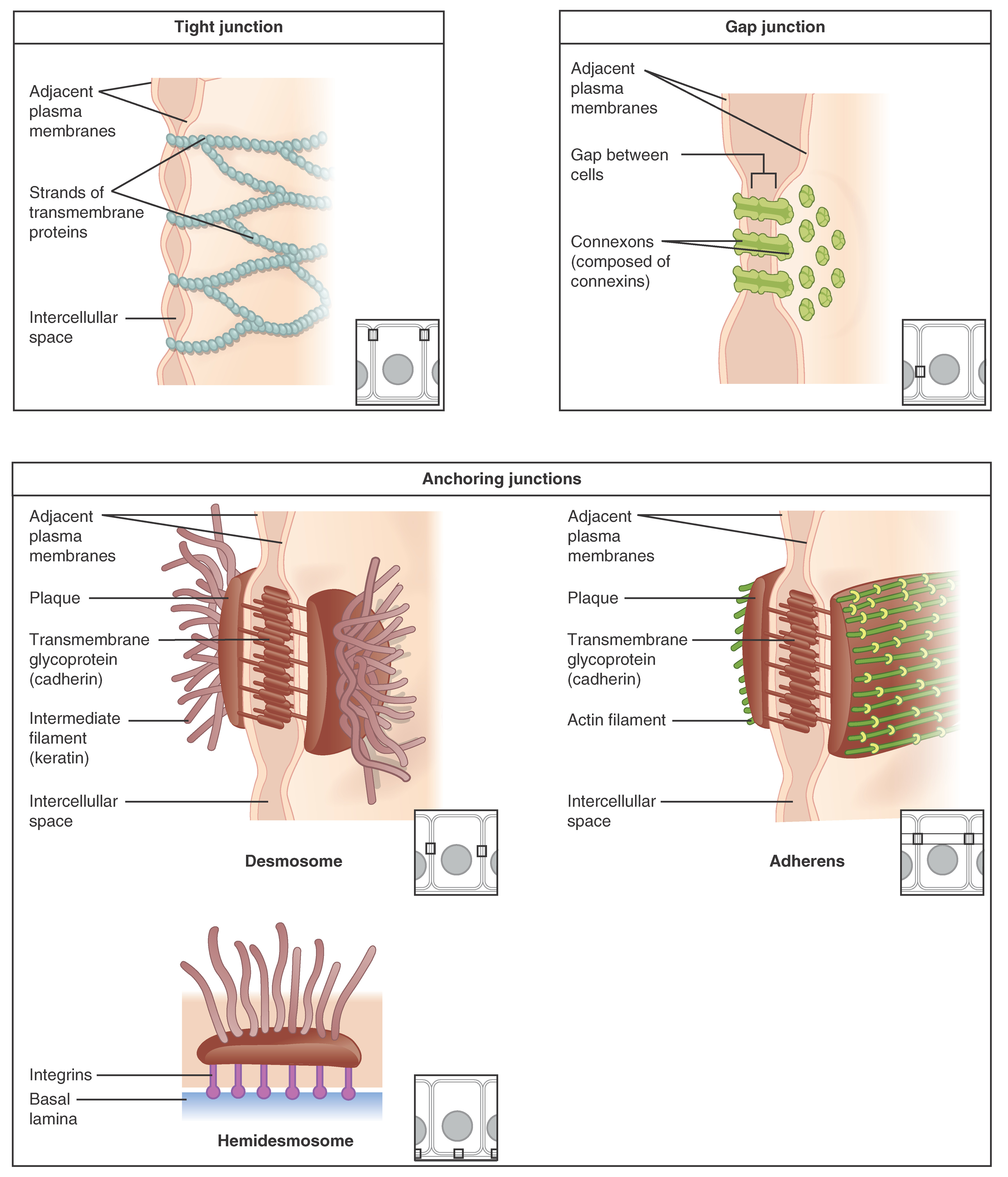
There are 3 types of cell to cell connections in epithelia. The tight junction    , which separates the cells into upper and lower compartments, which prevent the passage of materials through the cells. An anchoring junction    includes several types of cell junctions that help stabilize epithelial tissues. Desmosomes are common on surfaces of cells where they provide strong and flexible connections.

In contrast with the tight and anchoring junctions, a gap junction    forms an passageway between adjacent cells allowing the movement of small molecules and ions between the cells. These junctions coordinate the functions in large groups of cells.

Get Jobilize Job Search Mobile App in your pocket Now!

Get it on Google Play Download on the App Store Now


Source:  OpenStax, Histology. OpenStax CNX. Feb 27, 2015 Download for free at http://legacy.cnx.org/content/col11764/1.1
Google Play and the Google Play logo are trademarks of Google Inc.

Notification Switch

Would you like to follow the 'Histology' conversation and receive update notifications?

Ask