

| << Chapter < Page | Chapter >> Page > |
NOTE: Chapter Objectives: After studying the chapter, you will be able to:Describe the integumentary system and the role it plays in homeostasis. Describe the layers of the skin and the functions of each layer.Describe the accessory structures of the skin and the functions of each. Describe the changes that occur in the integumentary system during the aging process.Discuss several common diseases, disorders, and injuries that affect the integumentary system. Explain treatments for some common diseases, disorders, and injuries of the integumentary system.
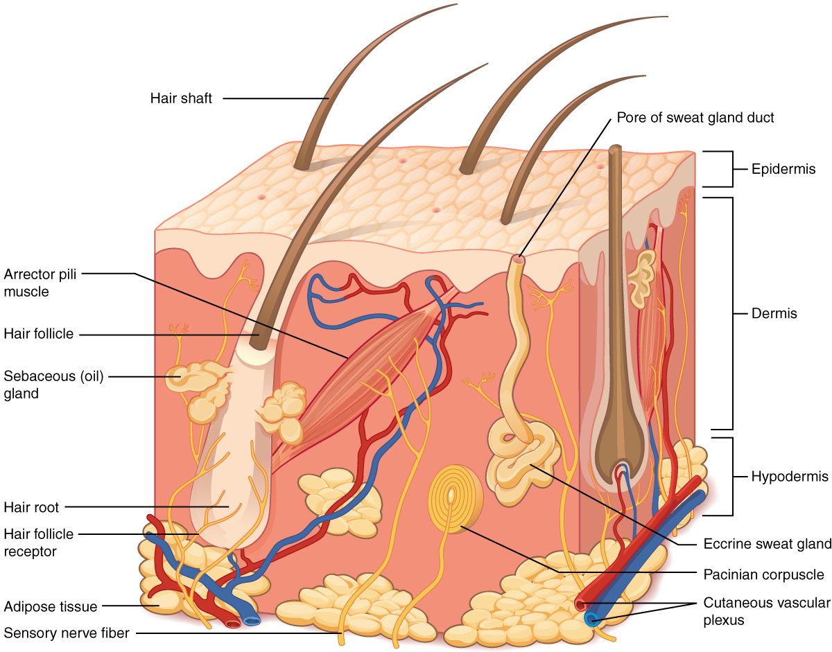
What do you think when you look at your skin in the mirror? Do you think about covering it with makeup, adding a tattoo, or maybe a body piercing? Or do you think about the fact that the skin belongs to one of the body’s most essential and dynamic systems: the integumentary system? The integumentary system refers to the skin and its accessory structures, and it is responsible for much more than simply lending to your outward appearance. In the adult human body, the skin makes up about 16 percent of body weight and covers an area of 1.5 to 2 m2. In fact, the skin and accessory structures are the largest organ system in the human body. As such, the skin protects your inner organs and it is in need of daily care and protection to maintain its health. This chapter will introduce the structure and functions of the integumentary system, as well as some of the diseases, disorders, and injuries that can affect this system. Although you may not typically think of the skin as an organ, it is in fact made of tissues that work together as a single structure to perform unique and critical functions. The skin and its accessory structures make up the integumentary system , which provides the body with overall protection. The skin is made of multiple layers of cells and tissues, which are held to underlying structures by connective tissue ( [link] ). The deeper layer of skin is well vascularized (has numerous blood vessels). It also has numerous sensory, and autonomic and sympathetic nerve fibers ensuring communication to and from the brain.
The epidermis is composed of keratinized, stratified squamous epithelium. It is made of four or five layers of epithelial cells, depending on its location in the body. It does not have any blood vessels within it. Tissue without its own blood supply is referred to as avascular . Skin that has four layers of cells is referred to as thin skin . From deep to superficial, these layers are the stratum basale, stratum spinosum, stratum granulosum, and stratum corneum. Most of the skin can be classified as thin skin. Thick skin is found only on the palms of the hands and the soles of the feet. It has a 5th layer called the stratum lucidum , located between the stratum corneum and the stratum granulosum ( [link] ).
Notification Switch
Would you like to follow the 'Integumentary system' conversation and receive update notifications?

|
|

|
|

|
|

|
|

|
|

|
|

|
|

|
|

|
|

|