<< Chapter < Page Chapter >> Page >
a) A micrograph of a spherical cell approximately 4 µm in diameter. B) A micrograph of wavy ribbon shaped cells approximately 10 µm in length. C) a micrograph of a bell shaped cell approximately 50µm in diameter with a tail approximately 200 µm in length. D) An oval shaped cell approximately 100 µm in length. A ring shaped cell approximately 4 µm in diameter; the ring shaped cell is inside a red blood cell.
Eukaryotic cells come in a variety of cell shapes. (a) Spheroid Chromulina alga. (b) Fusiform shaped Trypanosoma . (c) Bell-shaped Vorticella . (d) Ovoid Paramecium . (e) Ring-shaped Plasmodium ovale . (credit a: modification of work by NOAA; credit b, e: modification of work by Centers for Disease Control and Prevention)
  • Identify two differences between eukaryotic and prokaryotic cells.

Nucleus

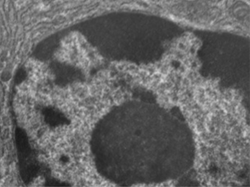
Unlike prokaryotic cells, in which DNA is loosely contained in the nucleoid region, eukaryotic cells possess a nucleus , which is surrounded by a complex nuclear membrane that houses the DNA genome ( [link] ). By containing the cell’s DNA, the nucleus ultimately controls all activities of the cell and also serves an essential role in reproduction and heredity. Eukaryotic cells typically have their DNA organized into multiple linear chromosomes. The DNA within the nucleus is highly organized and condensed to fit inside the nucleus, which is accomplished by wrapping the DNA around proteins called histones.

A micrograph of a portion of an oval cell. In the center is a darker spherical structure.
Eukaryotic cells have a well-defined nucleus. The nucleus of this mammalian lung cell is the large, dark, oval-shaped structure in the lower half of the image.

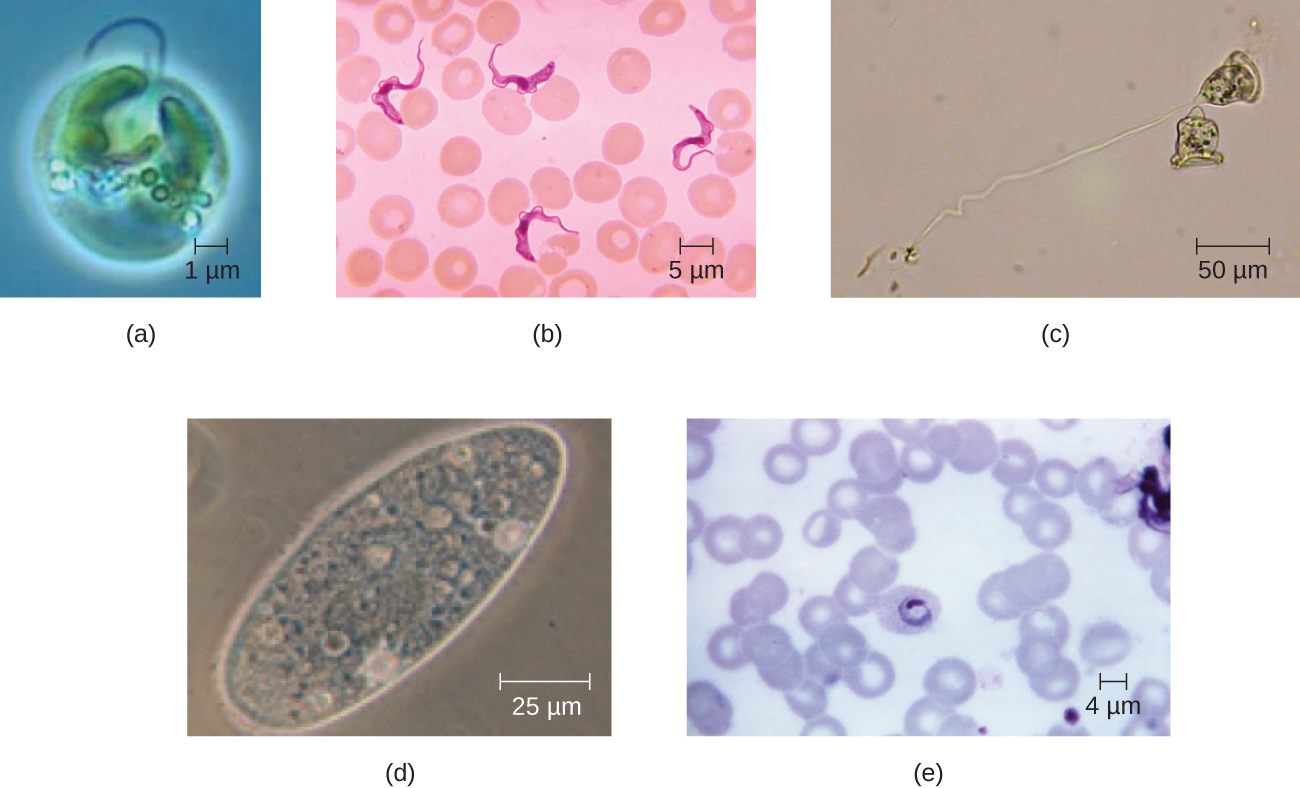
Although most eukaryotic cells have only one nucleus, exceptions exist. For example, protozoans of the genus Paramecium typically have two complete nuclei: a small nucleus that is used for reproduction (micronucleus) and a large nucleus that directs cellular metabolism (macronucleus). Additionally, some fungi transiently form cells with two nuclei, called heterokaryotic cells, during sexual reproduction. Cells whose nuclei divide, but whose cytoplasm does not, are called coenocytes .

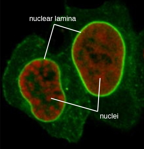
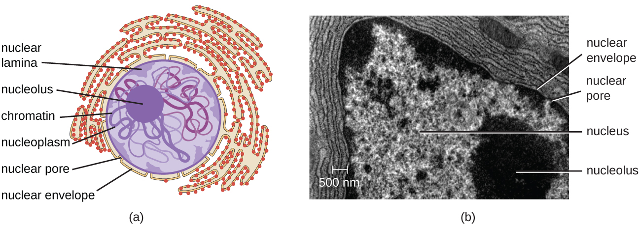
The nucleus is bound by a complex nuclear membrane , often called the nuclear envelope , that consists of two distinct lipid bilayers that are contiguous with each other ( [link] ). Despite these connections between the inner and outer membranes, each membrane contains unique lipids and proteins on its inner and outer surfaces. The nuclear envelope contains nuclear pores, which are large, rosette-shaped protein complexes that control the movement of materials into and out of the nucleus. The overall shape of the nucleus is determined by the nuclear lamina , a meshwork of intermediate filaments found just inside the nuclear envelope membranes. Outside the nucleus, additional intermediate filaments form a looser mesh and serve to anchor the nucleus in position within the cell.

A micrograph showing an oval cell with a large oval nucleus. The nucleus is red with a bright green outline labeled nuclear lamina. Green lines criss-cross the rest of the cell outside the nucleus.
In this fluorescent microscope image, all the intermediate filaments have been stained with a bright green fluorescent stain. The nuclear lamina is the intense bright green ring around the faint red nuclei.

Nucleolus

The nucleolus is a dense region within the nucleus where ribosomal RNA (rRNA) biosynthesis occurs. In addition, the nucleolus is also the site where assembly of ribosomes begins. Preribosomal complexes are assembled from rRNA and proteins in the nucleolus; they are then transported out to the cytoplasm, where ribosome assembly is completed ( [link] ).

a) A diagram showing the nucleus. A sphere in the center of the nucleus is labeled nucleolus. Lines within the nucleus are labeled chromatin. The fluid of the nucleus is labeled nucleoplasm. The outer region just inside the nuclear envelope is labeled nuclear lamina. The outside of the nucleus is labeled nuclear envelop and pores in the envelope are labeled nuclear pores.  The nuclear envelope is continuous with and becomes the endoplasmic reticulum; a webbing of membranes outside the nucleus. B) A micrograph showing these same structures. The nucleolus is a dark region inside the nucleus which is composed of many lighter lines. The nuclear envelop forms the outside of the nucleus and a pore is seen as a light region in the envelope. Outside the envelope are many lines labeled rough endoplasmic reticulum. A smaller set of lines is labeled mitochondrion overlaying part of the RER.
(a) The nucleolus is the dark, dense area within the nucleus. It is the site of rRNA synthesis and preribosomal assembly. (b) Electron micrograph showing the nucleolus.

Questions & Answers

what is biology
Hajah Reply
the study of living organisms and their interactions with one another and their environments
AI-Robot
what is biology
Victoria Reply
HOW CAN MAN ORGAN FUNCTION
Alfred Reply
the diagram of the digestive system
Assiatu Reply
allimentary cannel
Ogenrwot
How does twins formed
William Reply
They formed in two ways first when one sperm and one egg are splited by mitosis or two sperm and two eggs join together
Oluwatobi
what is genetics
Josephine Reply
Genetics is the study of heredity
Misack
how does twins formed?
Misack
What is manual
Hassan Reply
discuss biological phenomenon and provide pieces of evidence to show that it was responsible for the formation of eukaryotic organelles
Joseph Reply
what is biology
Yousuf Reply
the study of living organisms and their interactions with one another and their environment.
Wine
discuss the biological phenomenon and provide pieces of evidence to show that it was responsible for the formation of eukaryotic organelles in an essay form
Joseph Reply
what is the blood cells
Shaker Reply
list any five characteristics of the blood cells
Shaker
lack electricity and its more savely than electronic microscope because its naturally by using of light
Abdullahi Reply
advantage of electronic microscope is easily and clearly while disadvantage is dangerous because its electronic. advantage of light microscope is savely and naturally by sun while disadvantage is not easily,means its not sharp and not clear
Abdullahi
cell theory state that every organisms composed of one or more cell,cell is the basic unit of life
Abdullahi
is like gone fail us
DENG
cells is the basic structure and functions of all living things
Ramadan
What is classification
ISCONT Reply
is organisms that are similar into groups called tara
Yamosa
in what situation (s) would be the use of a scanning electron microscope be ideal and why?
Kenna Reply
A scanning electron microscope (SEM) is ideal for situations requiring high-resolution imaging of surfaces. It is commonly used in materials science, biology, and geology to examine the topography and composition of samples at a nanoscale level. SEM is particularly useful for studying fine details,
Hilary
cell is the building block of life.
Condoleezza Reply
Got questions? Join the online conversation and get instant answers!
Jobilize.com Reply

Get Jobilize Job Search Mobile App in your pocket Now!

Get it on Google Play Download on the App Store Now


Source:  OpenStax, Microbiology. OpenStax CNX. Nov 01, 2016 Download for free at http://cnx.org/content/col12087/1.4
Google Play and the Google Play logo are trademarks of Google Inc.

Notification Switch

Would you like to follow the 'Microbiology' conversation and receive update notifications?

Ask