<< Chapter < Page Chapter >> Page >
A micrograph shows purple circles and pink rods.
In this specimen, the gram-positive bacterium Staphylococcus aureus retains crystal violet dye even after the decolorizing agent is added. Gram-negative Escherichia coli , the most common Gram stain quality-control bacterium, is decolorized, and is only visible after the addition of the pink counterstain safranin. (credit: modification of work by Nina Parker)
  • Explain the role of Gram’s iodine in the Gram stain procedure.
  • Explain the role of alcohol in the Gram stain procedure.
  • What color are gram-positive and gram-negative cells, respectively, after the Gram stain procedure?

Part 3

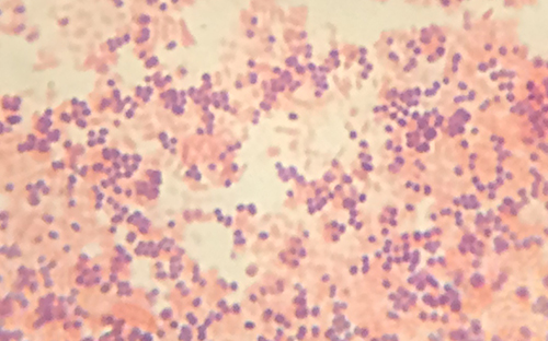
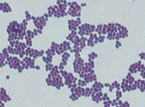
Viewing Cindy’s specimen under the darkfield microscope has provided the technician with some important clues about the identity of the microbe causing her infection. However, more information is needed to make a conclusive diagnosis. The technician decides to make a Gram stain of the specimen. This technique is commonly used as an early step in identifying pathogenic bacteria. After completing the Gram stain procedure , the technician views the slide under the brightfield microscope and sees purple, grape-like clusters of spherical cells ( [link] ).

  • Are these bacteria gram-positive or gram-negative?
  • What does this reveal about their cell walls?

Jump to the next Clinical Focus box. Go back to the previous Clinical Focus box.

A micrograph shows purple circles in random clusters.
(credit: modification of work by American Society for Microbiology)

Acid-fast stains

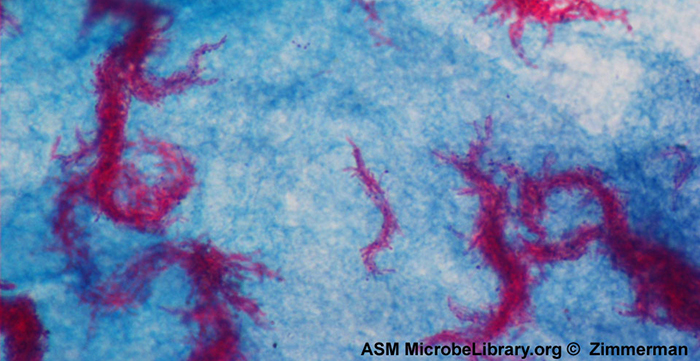
Acid-fast staining is another commonly used, differential staining technique that can be an important diagnostic tool. An acid-fast stain is able to differentiate two types of gram-positive cells: those that have waxy mycolic acids in their cell walls, and those that do not. Two different methods for acid-fast staining are the Ziehl-Neelsen technique and the Kinyoun technique . Both use carbolfuchsin as the primary stain. The waxy, acid-fast cells retain the carbolfuchsin even after a decolorizing agent (an acid-alcohol solution) is applied. A secondary counterstain, methylene blue, is then applied, which renders non–acid-fast cells blue.

The fundamental difference between the two carbolfuchsin-based methods is whether heat is used during the primary staining process. The Ziehl-Neelsen method uses heat to infuse the carbolfuchsin into the acid-fast cells, whereas the Kinyoun method does not use heat. Both techniques are important diagnostic tools because a number of specific diseases are caused by acid-fast bacteria (AFB). If AFB are present in a tissue sample, their red or pink color can be seen clearly against the blue background of the surrounding tissue cells ( [link] ).

  • Why are acid-fast stains useful?

Using microscopy to diagnose tuberculosis

Mycobacterium tuberculosis , the bacterium that causes tuberculosis , can be detected in specimens based on the presence of acid-fast bacilli. Often, a smear is prepared from a sample of the patient’s sputum and then stained using the Ziehl-Neelsen technique ( [link] ). If acid-fast bacteria are confirmed, they are generally cultured to make a positive identification. Variations of this approach can be used as a first step in determining whether M. tuberculosis or other acid-fast bacteria are present, though samples from elsewhere in the body (such as urine) may contain other Mycobacterium species.

An alternative approach for determining the presence of M. tuberculosis is immunofluorescence. In this technique, fluorochrome-labeled antibodies bind to M. tuberculosis , if present. Antibody-specific fluorescent dyes can be used to view the mycobacteria with a fluorescence microscope.

A micrograph shows red chains of cells on a blue background.
Ziehl-Neelsen staining has rendered these Mycobacterium tuberculosis cells red and the surrounding growth indicator medium blue. (credit: modification of work by American Society for Microbiology)

Questions & Answers

what is biology
Hajah Reply
the study of living organisms and their interactions with one another and their environments
AI-Robot
what is biology
Victoria Reply
HOW CAN MAN ORGAN FUNCTION
Alfred Reply
the diagram of the digestive system
Assiatu Reply
allimentary cannel
Ogenrwot
How does twins formed
William Reply
They formed in two ways first when one sperm and one egg are splited by mitosis or two sperm and two eggs join together
Oluwatobi
what is genetics
Josephine Reply
Genetics is the study of heredity
Misack
how does twins formed?
Misack
What is manual
Hassan Reply
discuss biological phenomenon and provide pieces of evidence to show that it was responsible for the formation of eukaryotic organelles
Joseph Reply
what is biology
Yousuf Reply
the study of living organisms and their interactions with one another and their environment.
Wine
discuss the biological phenomenon and provide pieces of evidence to show that it was responsible for the formation of eukaryotic organelles in an essay form
Joseph Reply
what is the blood cells
Shaker Reply
list any five characteristics of the blood cells
Shaker
lack electricity and its more savely than electronic microscope because its naturally by using of light
Abdullahi Reply
advantage of electronic microscope is easily and clearly while disadvantage is dangerous because its electronic. advantage of light microscope is savely and naturally by sun while disadvantage is not easily,means its not sharp and not clear
Abdullahi
cell theory state that every organisms composed of one or more cell,cell is the basic unit of life
Abdullahi
is like gone fail us
DENG
cells is the basic structure and functions of all living things
Ramadan
What is classification
ISCONT Reply
is organisms that are similar into groups called tara
Yamosa
in what situation (s) would be the use of a scanning electron microscope be ideal and why?
Kenna Reply
A scanning electron microscope (SEM) is ideal for situations requiring high-resolution imaging of surfaces. It is commonly used in materials science, biology, and geology to examine the topography and composition of samples at a nanoscale level. SEM is particularly useful for studying fine details,
Hilary
cell is the building block of life.
Condoleezza Reply
Got questions? Join the online conversation and get instant answers!
Jobilize.com Reply

Get Jobilize Job Search Mobile App in your pocket Now!

Get it on Google Play Download on the App Store Now


Source:  OpenStax, Microbiology. OpenStax CNX. Nov 01, 2016 Download for free at http://cnx.org/content/col12087/1.4
Google Play and the Google Play logo are trademarks of Google Inc.

Notification Switch

Would you like to follow the 'Microbiology' conversation and receive update notifications?

Ask