<< Chapter < Page
  Microbiology   Page 1 / 1
Chapter >> Page >
A micrograph showing spherical cells attached to a matrix on a surface. A photo of green water in a bucket.
Medical devices that are inserted into a patient’s body often become contaminated with a thin biofilm of microorganisms enmeshed in the sticky material they secrete. The electron micrograph (left) shows the inside walls of an in-dwelling catheter. Arrows point to the round cells of Staphylococcus aureus bacteria attached to the layers of extracellular substrate. The garbage can (right) served as a rain collector. The arrow points to a green biofilm on the sides of the container. (credit left: modification of work by Centers for Disease Control and Prevention; credit right: modification of work by NASA)

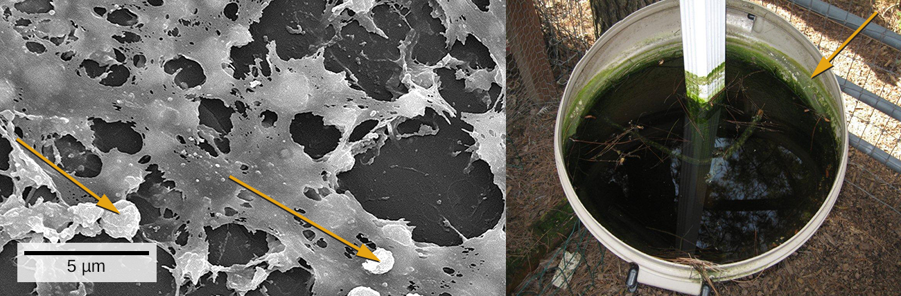
We are all familiar with the slimy layer on a pond surface or that makes rocks slippery. These are examples of biofilms—microorganisms embedded in thin layers of matrix material ( [link] ). Biofilms were long considered random assemblages of cells and had little attention from researchers. Recently, progress in visualization and biochemical methods has revealed that biofilms are an organized ecosystem within which many cells, usually of different species of bacteria, fungi, and algae, interact through cell signaling and coordinated responses. The biofilm provides a protected environment in harsh conditions and aids colonization by microorganisms. Biofilms also have clinical importance. They form on medical devices, resist routine cleaning and sterilization, and cause health-acquired infections. Within the body, biofilms form on the teeth as plaque, in the lungs of patients with cystic fibrosis, and on the cardiac tissue of patients with endocarditis. The slime layer helps protect the cells from host immune defenses and antibiotic treatments.

Studying biofilms requires new approaches. Because of the cells’ adhesion properties, many of the methods for culturing and counting cells that are explored in this chapter are not easily applied to biofilms. This is the beginning of a new era of challenges and rewarding insight into the ways that microorganisms grow and thrive in nature.

Questions & Answers

what is phylogeny
Odigie Reply
evolutionary history and relationship of an organism or group of organisms
AI-Robot
ok
Deng
what is biology
Hajah Reply
the study of living organisms and their interactions with one another and their environments
AI-Robot
what is biology
Victoria Reply
HOW CAN MAN ORGAN FUNCTION
Alfred Reply
the diagram of the digestive system
Assiatu Reply
allimentary cannel
Ogenrwot
How does twins formed
William Reply
They formed in two ways first when one sperm and one egg are splited by mitosis or two sperm and two eggs join together
Oluwatobi
what is genetics
Josephine Reply
Genetics is the study of heredity
Misack
how does twins formed?
Misack
What is manual
Hassan Reply
discuss biological phenomenon and provide pieces of evidence to show that it was responsible for the formation of eukaryotic organelles
Joseph Reply
what is biology
Yousuf Reply
the study of living organisms and their interactions with one another and their environment.
Wine
discuss the biological phenomenon and provide pieces of evidence to show that it was responsible for the formation of eukaryotic organelles in an essay form
Joseph Reply
what is the blood cells
Shaker Reply
list any five characteristics of the blood cells
Shaker
lack electricity and its more savely than electronic microscope because its naturally by using of light
Abdullahi Reply
advantage of electronic microscope is easily and clearly while disadvantage is dangerous because its electronic. advantage of light microscope is savely and naturally by sun while disadvantage is not easily,means its not sharp and not clear
Abdullahi
cell theory state that every organisms composed of one or more cell,cell is the basic unit of life
Abdullahi
is like gone fail us
DENG
cells is the basic structure and functions of all living things
Ramadan
What is classification
ISCONT Reply
is organisms that are similar into groups called tara
Yamosa
in what situation (s) would be the use of a scanning electron microscope be ideal and why?
Kenna Reply
A scanning electron microscope (SEM) is ideal for situations requiring high-resolution imaging of surfaces. It is commonly used in materials science, biology, and geology to examine the topography and composition of samples at a nanoscale level. SEM is particularly useful for studying fine details,
Hilary
Got questions? Join the online conversation and get instant answers!
Jobilize.com Reply

Get Jobilize Job Search Mobile App in your pocket Now!

Get it on Google Play Download on the App Store Now


Source:  OpenStax, Microbiology. OpenStax CNX. Nov 01, 2016 Download for free at http://cnx.org/content/col12087/1.4
Google Play and the Google Play logo are trademarks of Google Inc.

Notification Switch

Would you like to follow the 'Microbiology' conversation and receive update notifications?

Ask