<< Chapter < Page
  Microbiology   Page 1 / 1
Chapter >> Page >
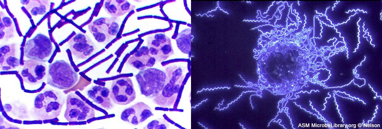
The left image shows a clear background with chains of solid purple rods and larger circular cells. The larger cells contain darker purple blotches inside each cell. The right image shows a black background with thin, glowing spirals.
Different types of microscopy are used to visualize different structures. Brightfield microscopy (left) renders a darker image on a lighter background, producing a clear image of these Bacillus anthracis cells in cerebrospinal fluid (the rod-shaped bacterial cells are surrounded by larger white blood cells). Darkfield microscopy (right) increases contrast, rendering a brighter image on a darker background, as demonstrated by this image of the bacterium Borrelia burgdorferi , which causes Lyme disease. (credit left: modification of work by Centers for Disease Control and Prevention; credit right: modification of work by American Society for Microbiology)

When we look at a rainbow, its colors span the full spectrum of light that the human eye can detect and differentiate. Each hue represents a different frequency of visible light, processed by our eyes and brains and rendered as red, orange, yellow, green, or one of the many other familiar colors that have always been a part of the human experience. But only recently have humans developed an understanding of the properties of light that allow us to see images in color.

Over the past several centuries, we have learned to manipulate light to peer into previously invisible worlds—those too small or too far away to be seen by the naked eye. Through a microscope, we can examine microbial cells and colonies, using various techniques to manipulate color, size, and contrast in ways that help us identify species and diagnose disease.

[link] illustrates how we can apply the properties of light to visualize and magnify images; but these stunning micrographs are just two examples of the numerous types of images we are now able to produce with different microscopic technologies. This chapter explores how various types of microscopes manipulate light in order to provide a window into the world of microorganisms. By understanding how various kinds of microscopes work, we can produce highly detailed images of microbes that can be useful for both research and clinical applications.

Questions & Answers

what is biology
Hajah Reply
the study of living organisms and their interactions with one another and their environments
AI-Robot
what is biology
Victoria Reply
HOW CAN MAN ORGAN FUNCTION
Alfred Reply
the diagram of the digestive system
Assiatu Reply
allimentary cannel
Ogenrwot
How does twins formed
William Reply
They formed in two ways first when one sperm and one egg are splited by mitosis or two sperm and two eggs join together
Oluwatobi
what is genetics
Josephine Reply
Genetics is the study of heredity
Misack
how does twins formed?
Misack
What is manual
Hassan Reply
discuss biological phenomenon and provide pieces of evidence to show that it was responsible for the formation of eukaryotic organelles
Joseph Reply
what is biology
Yousuf Reply
the study of living organisms and their interactions with one another and their environment.
Wine
discuss the biological phenomenon and provide pieces of evidence to show that it was responsible for the formation of eukaryotic organelles in an essay form
Joseph Reply
what is the blood cells
Shaker Reply
list any five characteristics of the blood cells
Shaker
lack electricity and its more savely than electronic microscope because its naturally by using of light
Abdullahi Reply
advantage of electronic microscope is easily and clearly while disadvantage is dangerous because its electronic. advantage of light microscope is savely and naturally by sun while disadvantage is not easily,means its not sharp and not clear
Abdullahi
cell theory state that every organisms composed of one or more cell,cell is the basic unit of life
Abdullahi
is like gone fail us
DENG
cells is the basic structure and functions of all living things
Ramadan
What is classification
ISCONT Reply
is organisms that are similar into groups called tara
Yamosa
in what situation (s) would be the use of a scanning electron microscope be ideal and why?
Kenna Reply
A scanning electron microscope (SEM) is ideal for situations requiring high-resolution imaging of surfaces. It is commonly used in materials science, biology, and geology to examine the topography and composition of samples at a nanoscale level. SEM is particularly useful for studying fine details,
Hilary
cell is the building block of life.
Condoleezza Reply
what is phylogeny
Odigie Reply
evolutionary history and relationship of an organism or group of organisms
AI-Robot
Got questions? Join the online conversation and get instant answers!
Jobilize.com Reply

Get Jobilize Job Search Mobile App in your pocket Now!

Get it on Google Play Download on the App Store Now


Source:  OpenStax, Microbiology. OpenStax CNX. Nov 01, 2016 Download for free at http://cnx.org/content/col12087/1.4
Google Play and the Google Play logo are trademarks of Google Inc.

Notification Switch

Would you like to follow the 'Microbiology' conversation and receive update notifications?

Ask