<< Chapter < Page Chapter >> Page >

Art connection

 Illustration depicts the embryo sac of an angiosperm, which is egg-shaped. The narrow end, called the micropylar end, has an opening that allows pollen to enter. The other end is called the chalazal end. Three cells called antipodals are at the chalazal end. The egg cell and two other cells called synergids are at the micropylar end. Two polar nuclei are inside the central cell in the middle of the embryo sac.
As shown in this diagram of the embryo sac in angiosperms, the ovule is covered by integuments and has an opening called a micropyle. Inside the embryo sac are three antipodal cells, two synergids, a central cell, and the egg cell.

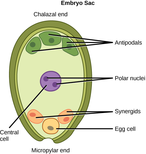
An embryo sac is missing the synergids. What specific impact would you expect this to have on fertilization?

  1. The pollen tube will be unable to form.
  2. The pollen tube will form but will not be guided toward the egg.
  3. Fertilization will not occur because the synergid is the egg.
  4. Fertilization will occur but the embryo will not be able to grow.

Sexual reproduction in gymnosperms

As with angiosperms, the lifecycle of a gymnosperm is also characterized by alternation of generations. In conifers such as pines, the green leafy part of the plant is the sporophyte, and the cones contain the male and female gametophytes ( [link] ). The female cones are larger than the male cones and are positioned towards the top of the tree; the small, male cones are located in the lower region of the tree. Because the pollen is shed and blown by the wind, this arrangement makes it difficult for a gymnosperm to self-pollinate.

 The conifer life cycle begins with a mature tree, which is called a sporophyte and is diploid (2n). The tree produces male cones in the lower branches, and female cones in the upper branches. The male cones produce pollen grains that contain two generative (sperm) nuclei and a tube nucleus. When the pollen lands on a female scale, a pollen tube grows toward the female gametophyte, which consists of an ovule containing the megaspore. Upon fertilization, a diploid zygote forms. The resulting seeds are dispersed, and grow into a mature tree, ending the cycle. Both the male and female cone are made up of rows of scales, but the male the female cone is round and wide, and the male cone is long and thin with thinner scales.
This image shows the life cycle of a conifer. Pollen from male cones blows up into upper branches, where it fertilizes female cones. Examples are shown of female and male cones. (credit “female”: modification of work by “Geographer”/Wikimedia Commons; credit “male”: modification of work by Roger Griffith)

Male gametophyte

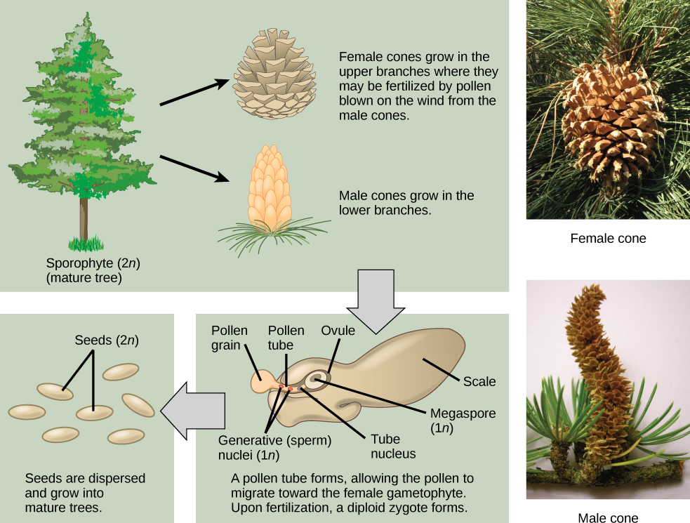
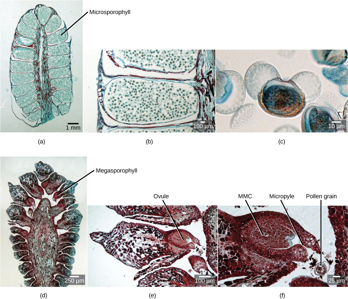
A male cone has a central axis on which bracts, a type of modified leaf, are attached. The bracts are known as microsporophylls ( [link] ) and are the sites where microspores will develop. The microspores develop inside the microsporangium. Within the microsporangium, cells known as microsporocytes divide by meiosis to produce four haploid microspores. Further mitosis of the microspore produces two nuclei: the generative nucleus, and the tube nucleus. Upon maturity, the male gametophyte (pollen) is released from the male cones and is carried by the wind to land on the female cone.

Watch this video to see a cedar releasing its pollen in the wind.

Female gametophyte

The female cone also has a central axis on which bracts known as megasporophylls ( [link] ) are present. In the female cone, megaspore mother cells are present in the megasporangium. The megaspore mother cell divides by meiosis to produce four haploid megaspores. One of the megaspores divides to form the multicellular female gametophyte, while the others divide to form the rest of the structure. The female gametophyte is contained within a structure called the archegonium.

 Part a shows a cross section of a male cone, which is oval with a flattened bottom. A stem-like structure runs up the middle, and oblong microsporophylls radiate from either side. Migrograph b shows a blowup of a microsphorphyll, which is filled with round pollen grains. Micrograph C shows a blowup of a pollen grain, which is oval with two lobes and resembles Mickey Mouse. Part D shows a cross section of a female cone, which is similar in shape to the male cone but with a wider central structure. Megasporophylls radiate from either side of this structure. At the base the megasprophylls are narrow, and the widen out into a roughly diamond shape. Part E shows a blowup of the megasprophyll, which has an oval ovule at its base. Part F shows a blowup of the ovule. The megaspore mother cell is at the center. An opening called a micropyle allows entry of the pollen tube from the base.
These series of micrographs shows male and female gymnosperm gametophytes. (a) This male cone, shown in cross section, has approximately 20 microsporophylls, each of which produces hundreds of male gametophytes (pollen grains). (b) Pollen grains are visible in this single microsporophyll. (c) This micrograph shows an individual pollen grain. (d) This cross section of a female cone shows portions of about 15 megasporophylls. (e) The ovule can be seen in this single megasporophyll. (f) Within this single ovule are the megaspore mother cell (MMC), micropyle, and a pollen grain. (credit: modification of work by Robert R. Wise; scale-bar data from Matt Russell)

Get Jobilize Job Search Mobile App in your pocket Now!

Get it on Google Play Download on the App Store Now


Source:  OpenStax, Biology. OpenStax CNX. Feb 29, 2016 Download for free at http://cnx.org/content/col11448/1.10
Google Play and the Google Play logo are trademarks of Google Inc.

Notification Switch

Would you like to follow the 'Biology' conversation and receive update notifications?

Ask