<< Chapter < Page Chapter >> Page >

Occipital bone

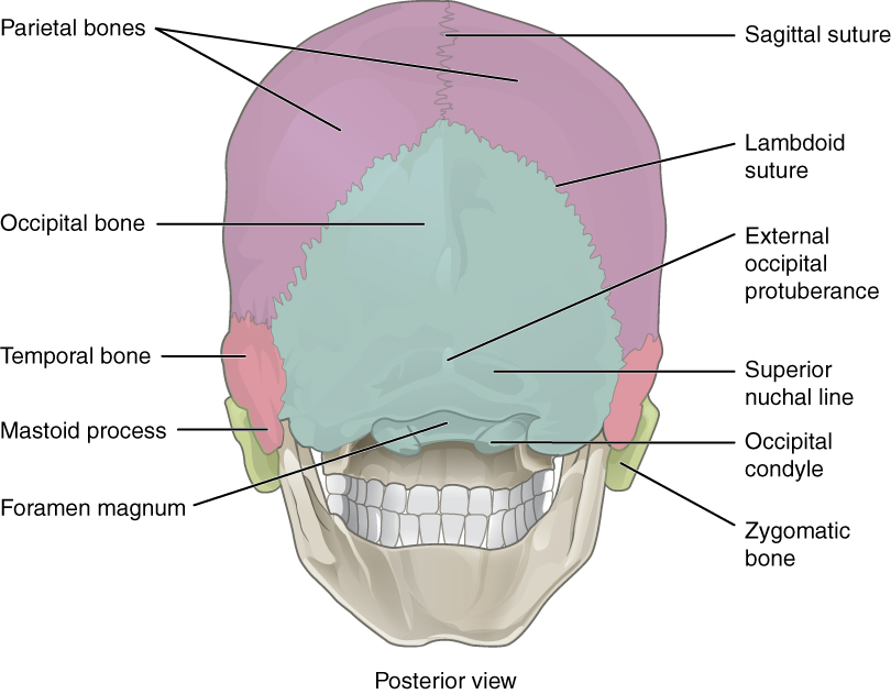
The occipital bone    is the single bone that forms the posterior skull and posterior base of the cranial cavity ( [link] ; see also [link] ). On the base of the skull, the occipital bone contains the large opening of the foramen magnum    , which allows for passage of the spinal cord as it exits the skull. On either side of the foramen magnum is an oval-shaped occipital condyle    . These condyles form joints with the first cervical vertebra and thus support the skull on top of the vertebral column.

Posterior view of skull

This figure shows the posterior view of the skull and the major parts are labeled.
This view of the posterior skull shows attachment sites for muscles and joints that support the skull.

Sphenoid bone

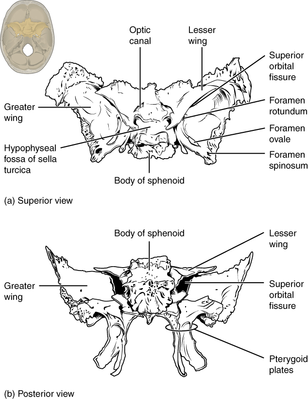
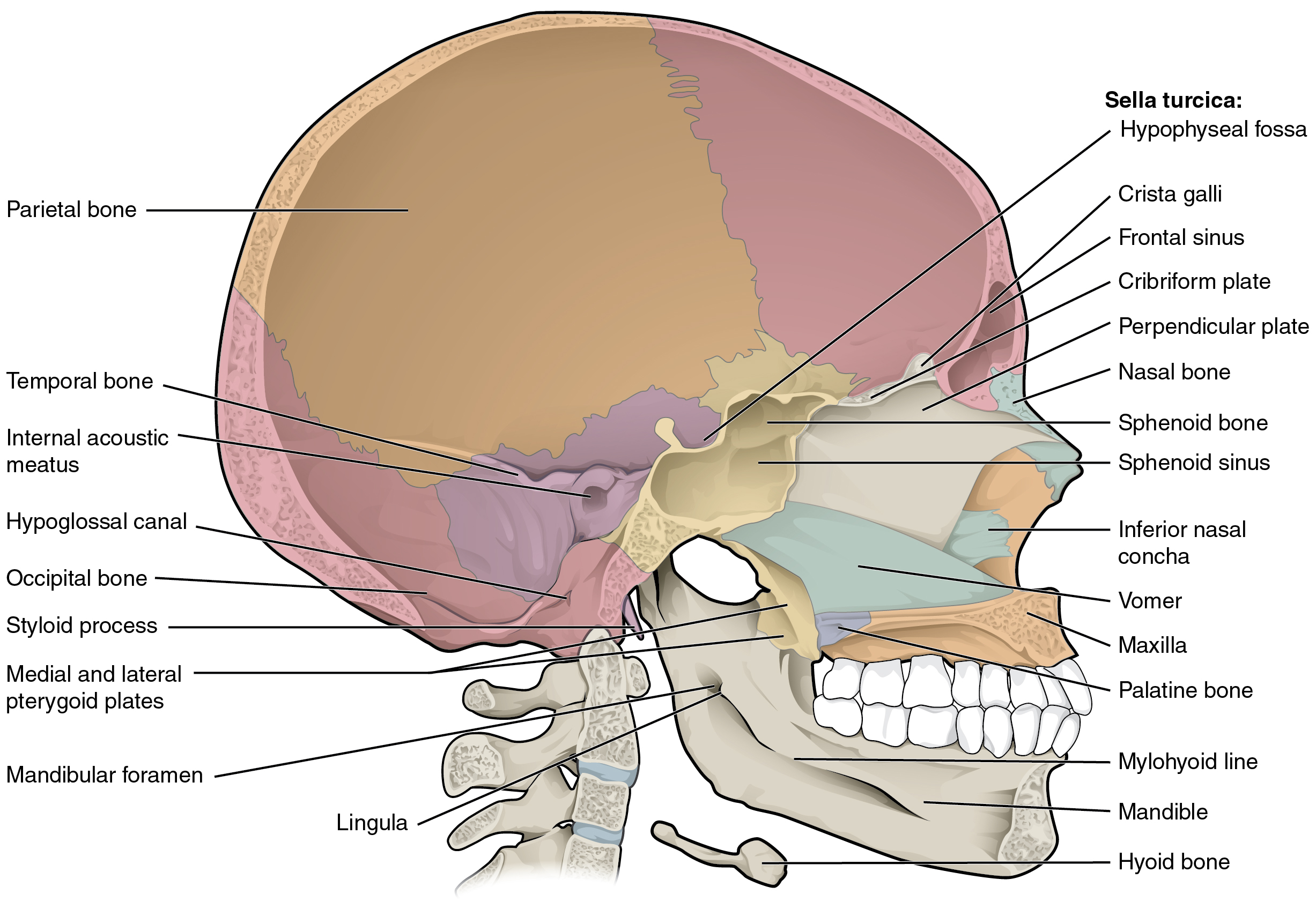
The sphenoid bone    is a single, complex bone of the central skull . It serves as a “keystone” bone, because it joins with almost every other bone of the skull. The sphenoid forms much of the base of the central skull and also extends to the temple area at the sides of the skull . Inside the cranial cavity, the bone with right and left wings which resemble the wings of a flying bird. The sella turcica    (“Turkish saddle”) is located at the midline of the sphenoid bone.

Sphenoid bone

This image shows the location and structure of the sphenoid bone. A small image of the skull on the top left shows the sphenoid bone highlighted in ochre yellow. The top panel shows the superior view of the sphenoid bone and the bottom panel shows the posterior view of the sphenoid bone.
Shown in isolation in (a) superior and (b) posterior views, the sphenoid bone is a single midline bone that forms the anterior walls and floor of the middle cranial fossa. It has a pair of lesser wings and a pair of greater wings. The sella turcica surrounds the hypophyseal fossa. Projecting downward are the medial and lateral pterygoid plates. The sphenoid has multiple openings for the passage of nerves and blood vessels, including the optic canal, superior orbital fissure, foramen rotundum, foramen ovale, and foramen spinosum.

Ethmoid bone

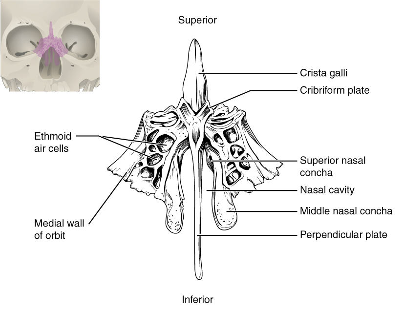
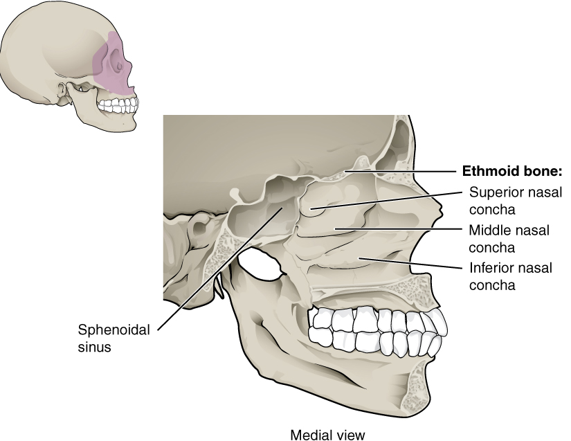
The ethmoid bone    is a single, midline bone that forms the roof and lateral walls of the upper nasal cavity, the upper portion of the nasal septum, and contributes to the medial wall of the orbit ( [link] and [link] ). On the interior of the skull, the ethmoid also forms a portion of the floor of the anterior cranial cavity (see [link] b ).

Sagittal section of skull

This diagram shows the sagittal section of the skull and identifies the major bones and cavities.
This midline view of the sagittally sectioned skull shows the nasal septum.

Ethmoid bone

This image shows the location and structure of the ethmoid bone. A small image of the skull on the top left shows the ethmoid bone colored in pink. A magnified image shows the inferior view of the ethmoid bone.
The unpaired ethmoid bone is located at the midline within the central skull. It has an upward projection, the crista galli, and a downward projection, the perpendicular plate, which forms the upper nasal septum. The cribriform plates form both the roof of the nasal cavity and a portion of the anterior cranial fossa floor. The lateral sides of the ethmoid bone form the lateral walls of the upper nasal cavity, part of the medial orbit wall, and give rise to the superior and middle nasal conchae. The ethmoid bone also contains the ethmoid air cells.

Lateral wall of nasal cavity

This figure shows the structure of the nasal cavity. A lateral view of the human skull is shown on the top left with the nasal cavity highlighted in purple. A magnified view of the nasal cavity shows the various bones present and their location.
The three nasal conchae are curved bones that project from the lateral walls of the nasal cavity. The superior nasal concha and middle nasal concha are parts of the ethmoid bone. The inferior nasal concha is an independent bone of the skull.

Sutures of the skull

A suture    is an immobile joint between adjacent bones of the skull. The narrow gap between the bones is filled with dense, fibrous connective tissue that unites the bones. The long sutures located between the bones of the brain case are not straight, but instead follow irregular, tightly twisting paths. These twisting lines serve to tightly interlock the adjacent bones, thus adding strength to the skull for brain protection.

Questions & Answers

Discuss the differences between taste and flavor, including how other sensory inputs contribute to our  perception of flavor.
John Reply
taste refers to your understanding of the flavor . while flavor one The other hand is refers to sort of just a blend things.
Faith
While taste primarily relies on our taste buds, flavor involves a complex interplay between taste and aroma
Kamara
which drugs can we use for ulcers
Ummi Reply
omeprazole
Kamara
what
Renee
what is this
Renee
is a drug
Kamara
of anti-ulcer
Kamara
Omeprazole Cimetidine / Tagament For the complicated once ulcer - kit
Patrick
what is the function of lymphatic system
Nency Reply
Not really sure
Eli
to drain extracellular fluid all over the body.
asegid
The lymphatic system plays several crucial roles in the human body, functioning as a key component of the immune system and contributing to the maintenance of fluid balance. Its main functions include: 1. Immune Response: The lymphatic system produces and transports lymphocytes, which are a type of
asegid
to transport fluids fats proteins and lymphocytes to the blood stream as lymph
Adama
what is anatomy
Oyindarmola Reply
Anatomy is the identification and description of the structures of living things
Kamara
what's the difference between anatomy and physiology
Oyerinde Reply
Anatomy is the study of the structure of the body, while physiology is the study of the function of the body. Anatomy looks at the body's organs and systems, while physiology looks at how those organs and systems work together to keep the body functioning.
AI-Robot
what is enzymes all about?
Mohammed Reply
Enzymes are proteins that help speed up chemical reactions in our bodies. Enzymes are essential for digestion, liver function and much more. Too much or too little of a certain enzyme can cause health problems
Kamara
yes
Prince
how does the stomach protect itself from the damaging effects of HCl
Wulku Reply
little girl okay how does the stomach protect itself from the damaging effect of HCL
Wulku
it is because of the enzyme that the stomach produce that help the stomach from the damaging effect of HCL
Kamara
function of digestive system
Ali Reply
function of digestive
Ali
the diagram of the lungs
Adaeze Reply
what is the normal body temperature
Diya Reply
37 degrees selcius
Xolo
37°c
Stephanie
please why 37 degree selcius normal temperature
Mark
36.5
Simon
37°c
Iyogho
the normal temperature is 37°c or 98.6 °Fahrenheit is important for maintaining the homeostasis in the body the body regular this temperature through the process called thermoregulation which involves brain skin muscle and other organ working together to maintain stable internal temperature
Stephanie
37A c
Wulku
what is anaemia
Diya Reply
anaemia is the decrease in RBC count hemoglobin count and PVC count
Eniola
what is the pH of the vagina
Diya Reply
how does Lysin attack pathogens
Diya
acid
Mary
I information on anatomy position and digestive system and there enzyme
Elisha Reply
anatomy of the female external genitalia
Muhammad Reply
Organ Systems Of The Human Body (Continued) Organ Systems Of The Human Body (Continued)
Theophilus Reply
what's lochia albra
Kizito
Got questions? Join the online conversation and get instant answers!
Jobilize.com Reply

Get Jobilize Job Search Mobile App in your pocket Now!

Get it on Google Play Download on the App Store Now


Source:  OpenStax, Skeletal system. OpenStax CNX. Apr 17, 2015 Download for free at https://legacy.cnx.org/content/col11779/1.1
Google Play and the Google Play logo are trademarks of Google Inc.

Notification Switch

Would you like to follow the 'Skeletal system' conversation and receive update notifications?

Ask