

| << Chapter < Page | Chapter >> Page > |
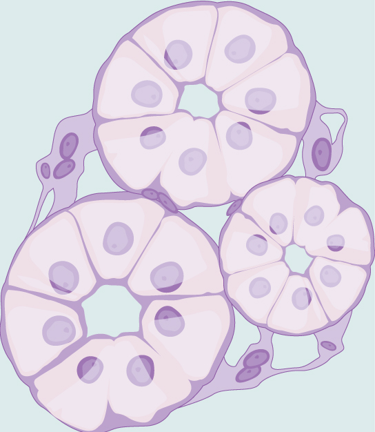
Cuboidal epithelial cells, shown in [link] , are cube-shaped with a single, central nucleus. They are most commonly found in a single layer representing a simple epithelia in glandular tissues (e.g. pancreas, mammary gland, etc.). They are also found in the walls of tubules and in the ducts of the kidney and liver.
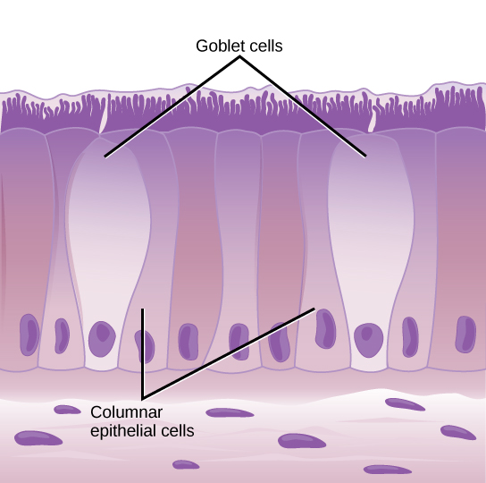
Columnar epithelial cells are taller than they are wide: they resemble a stack of columns in an epithelial layer, and are most commonly found in a single-layer arrangement. The nuclei of columnar epithelial cells in the digestive tract appear to be lined up at the base of the cells, as illustrated in [link] . These cells absorb material from the lumen of the digestive tract and prepare it for entry into the body through the circulatory and lymphatic systems.
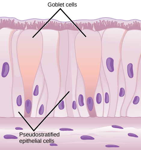
Columnar epithelial cells lining the respiratory tract appear to be stratified. However, each cell is attached to the base membrane of the tissue and, therefore, they are simple tissues. The nuclei are arranged at different levels in the layer of cells, making it appear as though there is more than one layer, as seen in [link] . This is called pseudostratified, columnar epithelia. This cellular covering has cilia at the apical, or free, surface of the cells. The cilia enhance the movement of mucous and trapped particles out of the respiratory tract, helping to protect the system from invasive microorganisms and harmful material that has been breathed into the body. Goblet cells are interspersed in some tissues (such as the lining of the trachea). The goblet cells contain mucous that traps irritants, which in the case of the trachea keep these irritants from getting into the lungs.
Connective tissues are made up of a matrix consisting of living cells and a non-living substance, called the ground substance. The ground substance is made of an organic substance (usually a protein) and an inorganic substance (usually a mineral or water). The principal cell of connective tissues is the fibroblast. This cell makes the fibers found in nearly all of the connective tissues. Fibroblasts are motile, able to carry out mitosis, and can synthesize whichever connective tissue is needed. Macrophages, lymphocytes, and, occasionally, leukocytes can be found in some connective tissues. Other cells that can be found in different connective tissues provide functions that are important for those specific tissues. For example, osteocytes (found in bone) and adipocytes (found in fat) are specialized cells that are critical for the formation and function of those tissues.
Notification Switch
Would you like to follow the 'Principles of biology' conversation and receive update notifications?

|
|

|
|

|
|

|
|

|
|

|
|

|
|

|
|

|
|

|