

|
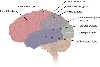
Ch 04: Spinal Cord General Anatomy & Brain Stem
Author:
M.D.Stephen VoronLecturer
University of Utah
USA
Access: |
1.1 An introduction to the human body Read Online
1.2 The chemical level of organization Read Online
After studying this chapter, you will be able to:
Though you may approach a course in anatomy and physiology strictly as a requirement for your field of study, the knowledge you gain in this course will serve you well in many aspects of your life. An understanding of anatomy and physiology is not only fundamental to any career in the health professions, but it can also benefit your own health. Familiarity with the human body can help you make healthful choices and prompt you to take appropriate action when signs of illness arise. Your knowledge in this field will help you understand news about nutrition, medications, medical devices, and procedures and help you understand genetic or infectious diseases. At some point, everyone will have a problem with some aspect of his or her body and your knowledge can help you to be a better parent, spouse, partner, friend, colleague, or caregiver.
This chapter begins with an overview of anatomy and physiology and a preview of the body regions and functions. It then covers the characteristics of life and how the body works to maintain stable conditions. It introduces a set of standard terms for body structures and for planes and positions in the body that will serve as a foundation for more comprehensive information covered later in the text. It ends with examples of medical imaging used to see inside the living body.
Question: White matter is a collection of myelinated and unmyelinated axons that conduct signals from one area of gray matter to another. What cell bodies can be recognized in white matter?
Choices:
Cell bodies of glial cells.
Cell bodies of ependymal cells.
Cell bodies of pial cells.
Cell bodies of neurons.
Question: The spinal nerves consist of ventral and dorsal roots. Where are the cell bodies of the axons in each root?
Choices:
Ventral and dorsal root cell bodies are in ganglia, (clusters of cell bodies outside the CNS).
Ventral and dorsal root cell bodies are in the gray matter of the cord.
Ventral root cell bodies are in the gray matter of the spinal cord and dorsal root cell bodies are in ganglia.
Ventral root cell bodies are in ganglia and dorsal root cell bodies are in the gray matter of the spinal cord.
Question: Consider this nucleus, where do its axons exit the medulla?
Choices:
Between the pons and medulla.
Laterally, between the ventral and dorsal roots, these axons are then known as "lateral roots''.
Between the two pyramids.
At the sulcus limitans.
Between the pyramid and the olive.
Question: Concerning spinal nerve C-8, which of the following is true?
Choices:
C-8 enters/exits between vertebrae C6 and C7.
C-8 enters/exits between vertebrae C7 and C8.
C-8 enters/exits between vertebrae T1 and T2.
C-8 enters/exits between vertebrae C7 and T1.
Question: Which of the following is/are true regarding this structure?
Choices:
It is attached to the conus medullaris.
It is a continuation of the pia and ependyma of the spinal cord.
It penetrates the dura at the end of the dural sac (vertebra S2).
It terminates as the coccygeal ligament fusing with the periosteum of the coccyx.
All of the above.
Question: What is true regarding the cervical (circle) and lumbar enlargements of the spinal cord? (Scroll down to see all choices).
Choices:
The extensive innervation required by neck structures, such as the larynx and pharynx, and by lower abdominal structures such as the bladder and reproductive organs cause an increase in gray and white matter.
The cord is larger because an increased number of axons and cell bodies is required in the cervical and lumbar regions to innervate the skin and muscles of the appendages.
There is an increase in white matter in the cervical region and an increase in gray matter in the lumbar region.
There is an increase in gray matter in the cervical region because of the increased motor innervation of the arms and an increase in white matter in the lumbar region because of the increased sensory innervation of the genitalia.
Question: What cord segments comprise the conus medullaris, and what do they innervate?
Choices:
The conus consists of lower sacral and a small coccygeal segment that innervates the perineum.
The conus consists of the lumbar and sacral segments which innervate the lower part of the body from the pelvis down.
The conus consists of S1-S5 and 3-4 coccygeal segments and innervates the pelvic area.
Question: The spinal pia forms collagenous ligaments that anchor it to the dura. These are the denticulate ligaments. What is their spatial relationship to the dorsal and ventral roots?
Choices:
The denticulate ligaments form a scalloped series of attachments between the ventral and dorsal roots in the cervical regions.
The denticulate ligaments are located below the dorsal roots on each side of the spinal cord.
The denticulate ligaments form a continuous sheet-like attachment above the dorsal roots.
The denticulate ligaments attach between the exits and entrances of the ventral and dorsal roots forming each spinal nerve.
Question: How do the meninges cover the spinal cord?
Choices:
The pia, arachnoid, and dura cover the spinal cord in tight, closely apposed layers.
The dura, arachnoid, and pia of the brain and spinal cord are continuous. All three layers loosely cover the spinal cord.
The dura, arachnoid, and pia all cover the spinal cord; the dura and arachnoid are tightly connected with each other.
Only the dura mater continues down from the brain to cover the spinal cord, protecting it from the surrounding bone.
Question: What does the cauda equina (Latin for horse's tail) represent?
Choices:
The ventral roots that extend from the lower spinal segments down to their various exits from the vertebral canal.
The dorsal roots that extend from their various entrances into the vertebral canal up to the proper segment of the cord.
Both dorsal and ventral roots within the subarachnoid space below the conus medullaris.
Question: If it were necessary, as it sometimes is, to insert a needle into the subarachnoid space to sample CSF, where is a relatively safe point for needle insertion?
Choices:
Between C-7 and T-1.
Between L-3 and L-4.
Between L-1 and L-2.
Between T-12 and L-1.

|
|

|
|

|
|

|
|
|
|
|

|
|

|
|

|
|

|
|

|
|

|
|

|