<< Chapter < Page Chapter >> Page >
In this illustration, the prokaryotic cell has an oval shape. The circular chromosome is concentrated in a region called the nucleoid. The fluid inside the cell is called the cytoplasm. Ribosomes, depicted as small dots, float in the cytoplasm. The cytoplasm is encased by a plasma membrane, which in turn is encased by a cell wall. A capsule surrounds the cell wall. The bacterium depicted has a flagellum protruding from one narrow end. Pili are small protrusions that project from the capsule in all directions.
The features of a typical bacterium cell are shown.

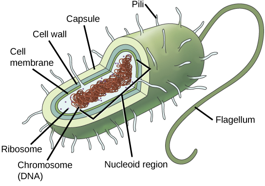
Both Bacteria and Archaea are types of prokaryotic cells. They differ in the lipid composition of their cell membranes and in the characteristics of their cell walls. Both types of prokaryotes have the same basic structures, but these are built from different chemical components that are evidence of an ancient separation of their lineages. The archaeal plasma membrane is chemically different from the bacterial membrane; some archaeal membranes are lipid monolayers instead of phosopholipid bilayers.

The cell wall

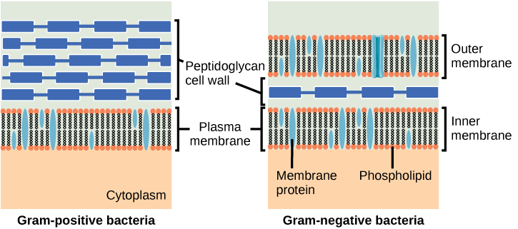
The cell wall is a protective layer that surrounds some prokaryotic cells and gives them shape and rigidity. It is located outside the cell membrane and prevents osmotic lysis (bursting caused by increasing volume). The chemical compositions of the cell walls vary between Archaea and Bacteria, as well as between bacterial species. Bacterial cell walls contain peptidoglycan    , composed of polysaccharide chains cross-linked to peptides. Bacteria are divided into two major groups: Gram-positive    and Gram-negative    , based on their reaction to a procedure called Gram staining. The different bacterial responses to the staining procedure are caused by cell wall structure. Gram-positive organisms have a thick wall consisting of many layers of peptidoglycan. Gram-negative bacteria have a thinner cell wall composed of a few layers of peptidoglycan and additional structures, surrounded by an outer membrane ( [link] ).

Art connection

This illustration compares Gram-positive to Gram-negative bacterial cell walls. The Gram-positive image on the left shows, from bottom to top: the cytoplasm, a plasma membrane bilayer with phospholipids and membrane proteins, and a thick cell wall with several layers of peptidoglycans. The Gram-negative image on the right shows, from bottom to top: the cytoplasm, a plasma membrane bilayer with phospholipids and membrane proteins, a thin cell wall with one layer of peptidoglycans, and an outer plasma membrane bilayer.
Bacteria are divided into two major groups: Gram-positive and Gram-negative. Both groups have a cell wall composed of peptidoglycans: In Gram-positive bacteria, the wall is thick, whereas in Gram-negative bacteria, the wall is thin. In Gram-negative bacteria, the cell wall is surrounded by an outer membrane.

Which of the following statements is true?

  1. Gram-positive bacteria have a single cell wall formed from peptidoglycan.
  2. Gram-positive bacteria have an outer membrane.
  3. The cell wall of Gram-negative bacteria is thick, and the cell wall of Gram-positive bacteria is thin.
  4. Gram-negative bacteria have a cell wall made of peptidoglycan, while Gram-positive bacteria have a cell wall made of phospholipids.

Archaeal cell walls do not contain peptidoglycan. There are four different types of archaeal cell walls. One type is composed of pseudopeptidoglycan    . The other three types of cell walls contain polysaccharides, glycoproteins, and surface-layer proteins known as S-layers.

Reproduction

Reproduction in prokaryotes is primarily asexual and takes place by binary fission. Recall that the DNA of a prokaryote exists usually as a single, circular chromosome. Prokaryotes do not undergo mitosis. Rather, the chromosome loop is replicated, and the two resulting copies attached to the plasma membrane move apart as the cell grows in a process called binary fission. The prokaryote, now enlarged, is pinched inward at its equator, and the two resulting cells, which are clones, separate. Binary fission does not provide an opportunity for genetic recombination, but prokaryotes can alter their genetic makeup in three ways.

Questions & Answers

what is phylogeny
Odigie Reply
evolutionary history and relationship of an organism or group of organisms
AI-Robot
ok
Deng
what is biology
Hajah Reply
the study of living organisms and their interactions with one another and their environments
AI-Robot
what is biology
Victoria Reply
HOW CAN MAN ORGAN FUNCTION
Alfred Reply
the diagram of the digestive system
Assiatu Reply
allimentary cannel
Ogenrwot
How does twins formed
William Reply
They formed in two ways first when one sperm and one egg are splited by mitosis or two sperm and two eggs join together
Oluwatobi
what is genetics
Josephine Reply
Genetics is the study of heredity
Misack
how does twins formed?
Misack
What is manual
Hassan Reply
discuss biological phenomenon and provide pieces of evidence to show that it was responsible for the formation of eukaryotic organelles
Joseph Reply
what is biology
Yousuf Reply
the study of living organisms and their interactions with one another and their environment.
Wine
discuss the biological phenomenon and provide pieces of evidence to show that it was responsible for the formation of eukaryotic organelles in an essay form
Joseph Reply
what is the blood cells
Shaker Reply
list any five characteristics of the blood cells
Shaker
lack electricity and its more savely than electronic microscope because its naturally by using of light
Abdullahi Reply
advantage of electronic microscope is easily and clearly while disadvantage is dangerous because its electronic. advantage of light microscope is savely and naturally by sun while disadvantage is not easily,means its not sharp and not clear
Abdullahi
cell theory state that every organisms composed of one or more cell,cell is the basic unit of life
Abdullahi
is like gone fail us
DENG
cells is the basic structure and functions of all living things
Ramadan
What is classification
ISCONT Reply
is organisms that are similar into groups called tara
Yamosa
in what situation (s) would be the use of a scanning electron microscope be ideal and why?
Kenna Reply
A scanning electron microscope (SEM) is ideal for situations requiring high-resolution imaging of surfaces. It is commonly used in materials science, biology, and geology to examine the topography and composition of samples at a nanoscale level. SEM is particularly useful for studying fine details,
Hilary
Got questions? Join the online conversation and get instant answers!
Jobilize.com Reply

Get Jobilize Job Search Mobile App in your pocket Now!

Get it on Google Play Download on the App Store Now


Source:  OpenStax, Concepts of biology. OpenStax CNX. Feb 29, 2016 Download for free at http://cnx.org/content/col11487/1.9
Google Play and the Google Play logo are trademarks of Google Inc.

Notification Switch

Would you like to follow the 'Concepts of biology' conversation and receive update notifications?

Ask