<< Chapter < Page Chapter >> Page >
Illustration shows the major human blood vessels. From the heart, blood is pumped into the aorta and distributed to systemic arteries. The carotid arteries bring blood to the head. The brachial arteries bring blood to the arms. The thoracic aorta brings blood down the trunk of the body along the spine. The hepatic, gastric and renal arteries, which branch from the thoracic aorta, bring blood to the liver, stomach and kidneys, respectively. The iliac artery brings blood to the legs. Blood is returned to the heart through two major veins, the superior vena cava at the top, and the inferior vena cava at the bottom. The jugular veins return blood from the head. The basilic veins return blood from the arms.  The hepatic, gastric and renal veins return blood from the liver, stomach and kidneys, respectively. The iliac vein returns blood from the legs.
The major human arteries and veins are shown. (credit: modification of work by Mariana Ruiz Villareal)

Arterioles diverge into capillary beds. Capillary beds contain a large number (10 to 100) of capillaries that branch among the cells and tissues of the body. Capillaries are narrow-diameter tubes that can fit red blood cells through in single file and are the sites for the exchange of nutrients, waste, and oxygen with tissues at the cellular level. Fluid also crosses into the interstitial space from the capillaries. The capillaries converge again into venules that connect to minor veins that finally connect to major veins that take blood high in carbon dioxide back to the heart. Veins are blood vessels that bring blood back to the heart. The major veins drain blood from the same organs and limbs that the major arteries supply. Fluid is also brought back to the heart via the lymphatic system.

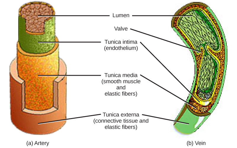
The structure of the different types of blood vessels reflects their function or layers. There are three distinct layers, or tunics, that form the walls of blood vessels ( [link] ). The first tunic is a smooth, inner lining of endothelial cells that are in contact with the red blood cells. The endothelial tunic is continuous with the endocardium of the heart. In capillaries, this single layer of cells is the location of diffusion of oxygen and carbon dioxide between the endothelial cells and red blood cells, as well as the exchange site via endocytosis and exocytosis. The movement of materials at the site of capillaries is regulated by vasoconstriction    , narrowing of the blood vessels, and vasodilation    , widening of the blood vessels; this is important in the overall regulation of blood pressure.

Veins and arteries both have two further tunics that surround the endothelium: the middle tunic is composed of smooth muscle and the outermost layer is connective tissue (collagen and elastic fibers). The elastic connective tissue stretches and supports the blood vessels, and the smooth muscle layer helps regulate blood flow by altering vascular resistance through vasoconstriction and vasodilation. The arteries have thicker smooth muscle and connective tissue than the veins to accommodate the higher pressure and speed of freshly pumped blood. The veins are thinner walled as the pressure and rate of flow are much lower. In addition, veins are structurally different than arteries in that veins have valves to prevent the backflow of blood. Because veins have to work against gravity to get blood back to the heart, contraction of skeletal muscle assists with the flow of blood back to the heart.

 Illustrations A and B show that arteries and veins consist of three layers, an inner endothelium called the tunica intima, a middle layer of smooth muscle and elastic fibers called the tunica media, and an outer layer of connective tissues and elastic fibers called the tunica externa. The outer two layers are thinner in the vein than in the artery. The central cavity is called the lumen. Veins have valves that extend into the lumen.
Arteries and veins consist of three layers: an outer tunica externa, a middle tunica media, and an inner tunica intima. Capillaries consist of a single layer of epithelial cells, the tunica intima. (credit: modification of work by NCI, NIH)

Section summary

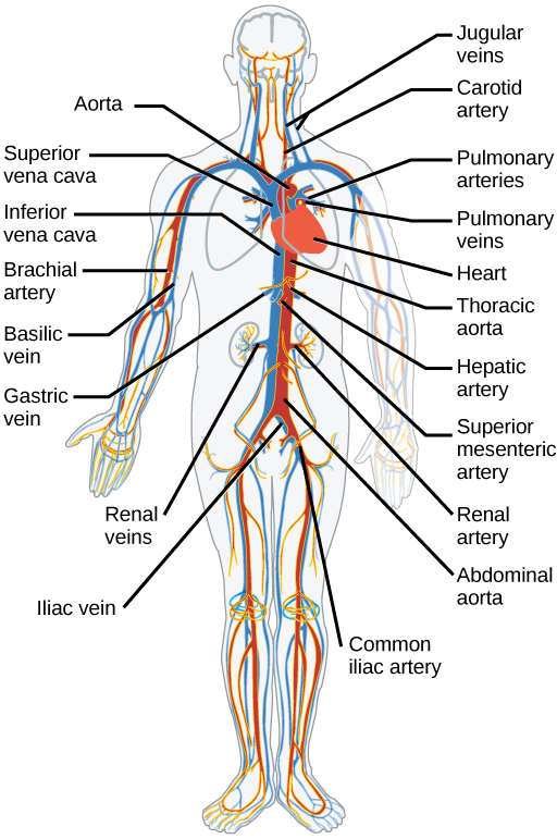
The heart muscle pumps blood through three divisions of the circulatory system: coronary, pulmonary, and systemic. There is one atrium and one ventricle on the right side and one atrium and one ventricle on the left side. The pumping of the heart is a function of cardiomyocytes, distinctive muscle cells that are striated like skeletal muscle but pump rhythmically and involuntarily like smooth muscle. The internal pacemaker starts at the sinoatrial node, which is located near the wall of the right atrium. Electrical charges pulse from the SA node causing the two atria to contract in unison; then the pulse reaches the atrioventricular node between the right atrium and right ventricle. A pause in the electric signal allows the atria to empty completely into the ventricles before the ventricles pump out the blood. The blood from the heart is carried through the body by a complex network of blood vessels; arteries take blood away from the heart, and veins bring blood back to the heart.

Art connections

[link] Which of the following statements about the circulatory system is false?

  1. Blood in the pulmonary vein is deoxygenated.
  2. Blood in the inferior vena cava is deoxygenated.
  3. Blood in the pulmonary artery is deoxygenated.
  4. Blood in the aorta is oxygenated.

[link] C

Got questions? Get instant answers now!

[link] Which of the following statements about the heart is false?

  1. The mitral valve separates the left ventricle from the left atrium.
  2. Blood travels through the bicuspid valve to the left atrium.
  3. Both the aortic and the pulmonary valves are semilunar valves.
  4. The mitral valve is an atrioventricular valve.

[link] B

Got questions? Get instant answers now!

Questions & Answers

calculate molarity of NaOH solution when 25.0ml of NaOH titrated with 27.2ml of 0.2m H2SO4
Gasin Reply
what's Thermochemistry
rhoda Reply
the study of the heat energy which is associated with chemical reactions
Kaddija
How was CH4 and o2 was able to produce (Co2)and (H2o
Edafe Reply
explain please
Victory
First twenty elements with their valences
Martine Reply
what is chemistry
asue Reply
what is atom
asue
what is the best way to define periodic table for jamb
Damilola Reply
what is the change of matter from one state to another
Elijah Reply
what is isolation of organic compounds
IKyernum Reply
what is atomic radius
ThankGod Reply
Read Chapter 6, section 5
Dr
Read Chapter 6, section 5
Kareem
Atomic radius is the radius of the atom and is also called the orbital radius
Kareem
atomic radius is the distance between the nucleus of an atom and its valence shell
Amos
Read Chapter 6, section 5
paulino
Bohr's model of the theory atom
Ayom Reply
is there a question?
Dr
when a gas is compressed why it becomes hot?
ATOMIC
It has no oxygen then
Goldyei
read the chapter on thermochemistry...the sections on "PV" work and the First Law of Thermodynamics should help..
Dr
Which element react with water
Mukthar Reply
Mgo
Ibeh
an increase in the pressure of a gas results in the decrease of its
Valentina Reply
definition of the periodic table
Cosmos Reply
What is the lkenes
Da Reply
what were atoms composed of?
Moses Reply
Got questions? Join the online conversation and get instant answers!
Jobilize.com Reply

Get Jobilize Job Search Mobile App in your pocket Now!

Get it on Google Play Download on the App Store Now


Source:  OpenStax, Biology. OpenStax CNX. Feb 29, 2016 Download for free at http://cnx.org/content/col11448/1.10
Google Play and the Google Play logo are trademarks of Google Inc.

Notification Switch

Would you like to follow the 'Biology' conversation and receive update notifications?

Ask