<< Chapter < Page Chapter >> Page >

Art connection

The kidney is shaped like a kidney bean standing on end. Two layers, the outer renal fascia and an inner capsule, cover the outside of the kidney. The inside of the kidney consists of three layers: the outer cortex, the middle medulla and the inner renal pelvis. The renal pelvis is flush with the concave side of the kidney, and empties into the ureter, a tube that runs down outside the concave side of the kidney. Nine renal pyramids are embedded in the medulla, which is the thickest kidney layer. Each renal pyramid is teardrop-shaped, with the narrow end facing the renal pelvis. The renal artery and renal vein enter the concave part of the kidney, just above the ureter. The renal artery and renal vein branch into arterioles and venuoles, respectively, which extend into the kidney and branch into capillaries in the cortex.
The internal structure of the kidney is shown. (credit: modification of work by NCI)

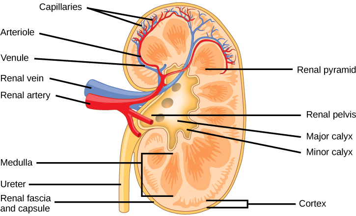
Which of the following statements about the kidney is false?

  1. The renal pelvis drains into the ureter.
  2. The renal pyramids are in the medulla.
  3. The cortex covers the capsule.
  4. Nephrons are in the renal cortex.

Because the kidney filters blood, its network of blood vessels is an important component of its structure and function. The arteries, veins, and nerves that supply the kidney enter and exit at the renal hilum. Renal blood supply starts with the branching of the aorta into the renal arteries (which are each named based on the region of the kidney they pass through) and ends with the exiting of the renal veins to join the inferior vena cava    . The renal arteries split into several segmental arteries upon entering the kidneys. Each segmental artery splits further into several interlobar arteries and enters the renal columns, which supply the renal lobes. The interlobar arteries split at the junction of the renal cortex and medulla to form the arcuate arteries . The arcuate “bow shaped” arteries form arcs along the base of the medullary pyramids. Cortical radiate arteries , as the name suggests, radiate out from the arcuate arteries. The cortical radiate arteries branch into numerous afferent arterioles, and then enter the capillaries supplying the nephrons. Veins trace the path of the arteries and have similar names, except there are no segmental veins.

As mentioned previously, the functional unit of the kidney is the nephron, illustrated in [link] . Each kidney is made up of over one million nephrons that dot the renal cortex, giving it a granular appearance when sectioned sagittally. There are two types of nephrons— cortical nephrons (85 percent), which are deep in the renal cortex, and juxtamedullary nephrons (15 percent), which lie in the renal cortex close to the renal medulla. A nephron consists of three parts—a renal corpuscle    , a renal tubule    , and the associated capillary network, which originates from the cortical radiate arteries.

Art connection

Illustration shows the nephron, a tube-like structure that begins in the kidney cortex. Here, arterioles converge in a bulb-like structure called the glomerulus, which is partly surrounded by a Bowman’s capsule. Afferent arterioles enter the glomerulus, and efferent arterioles leave. The glomerulus empties into the proximal convoluted tubule. A long loop, called the loop of Henle, extends from the proximal convoluted tubule to the inner medulla of the kidney, and then back out to the cortex. There, the loop of Henle joins a distal convoluted tubule. The distal convoluted tubule joins a collecting duct, which travels from the medulla back into the cortex, toward the center of the kidney. Eventually, the contents of the renal pyramid empty into the renal pelvis, and then the ureter.
The nephron is the functional unit of the kidney. The glomerulus and convoluted tubules are located in the kidney cortex, while collecting ducts are located in the pyramids of the medulla. (credit: modification of work by NIDDK)

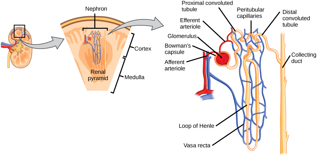
Which of the following statements about the nephron is false?

  1. The collecting duct empties into the distal convoluted tubule.
  2. The Bowman’s capsule surrounds the glomerulus.
  3. The loop of Henle is between the proximal and distal convoluted tubules.
  4. The loop of Henle empties into the distal convoluted tubule.

Renal corpuscle

The renal corpuscle, located in the renal cortex, is made up of a network of capillaries known as the glomerulus    and the capsule, a cup-shaped chamber that surrounds it, called the glomerular or Bowman's capsule    .

Renal tubule

The renal tubule is a long and convoluted structure that emerges from the glomerulus and can be divided into three parts based on function. The first part is called the proximal convoluted tubule (PCT)    due to its proximity to the glomerulus; it stays in the renal cortex. The second part is called the loop of Henle    , or nephritic loop, because it forms a loop (with descending and ascending limbs ) that goes through the renal medulla. The third part of the renal tubule is called the distal convoluted tubule (DCT)    and this part is also restricted to the renal cortex. The DCT, which is the last part of the nephron, connects and empties its contents into collecting ducts that line the medullary pyramids. The collecting ducts amass contents from multiple nephrons and fuse together as they enter the papillae of the renal medulla.

Questions & Answers

Discuss the differences between taste and flavor, including how other sensory inputs contribute to our  perception of flavor.
John Reply
taste refers to your understanding of the flavor . while flavor one The other hand is refers to sort of just a blend things.
Faith
While taste primarily relies on our taste buds, flavor involves a complex interplay between taste and aroma
Kamara
which drugs can we use for ulcers
Ummi Reply
omeprazole
Kamara
what
Renee
what is this
Renee
is a drug
Kamara
of anti-ulcer
Kamara
Omeprazole Cimetidine / Tagament For the complicated once ulcer - kit
Patrick
what is the function of lymphatic system
Nency Reply
Not really sure
Eli
to drain extracellular fluid all over the body.
asegid
The lymphatic system plays several crucial roles in the human body, functioning as a key component of the immune system and contributing to the maintenance of fluid balance. Its main functions include: 1. Immune Response: The lymphatic system produces and transports lymphocytes, which are a type of
asegid
to transport fluids fats proteins and lymphocytes to the blood stream as lymph
Adama
what is anatomy
Oyindarmola Reply
Anatomy is the identification and description of the structures of living things
Kamara
what's the difference between anatomy and physiology
Oyerinde Reply
Anatomy is the study of the structure of the body, while physiology is the study of the function of the body. Anatomy looks at the body's organs and systems, while physiology looks at how those organs and systems work together to keep the body functioning.
AI-Robot
what is enzymes all about?
Mohammed Reply
Enzymes are proteins that help speed up chemical reactions in our bodies. Enzymes are essential for digestion, liver function and much more. Too much or too little of a certain enzyme can cause health problems
Kamara
yes
Prince
how does the stomach protect itself from the damaging effects of HCl
Wulku Reply
little girl okay how does the stomach protect itself from the damaging effect of HCL
Wulku
it is because of the enzyme that the stomach produce that help the stomach from the damaging effect of HCL
Kamara
function of digestive system
Ali Reply
function of digestive
Ali
the diagram of the lungs
Adaeze Reply
what is the normal body temperature
Diya Reply
37 degrees selcius
Xolo
37°c
Stephanie
please why 37 degree selcius normal temperature
Mark
36.5
Simon
37°c
Iyogho
the normal temperature is 37°c or 98.6 °Fahrenheit is important for maintaining the homeostasis in the body the body regular this temperature through the process called thermoregulation which involves brain skin muscle and other organ working together to maintain stable internal temperature
Stephanie
37A c
Wulku
what is anaemia
Diya Reply
anaemia is the decrease in RBC count hemoglobin count and PVC count
Eniola
what is the pH of the vagina
Diya Reply
how does Lysin attack pathogens
Diya
acid
Mary
I information on anatomy position and digestive system and there enzyme
Elisha Reply
anatomy of the female external genitalia
Muhammad Reply
Organ Systems Of The Human Body (Continued) Organ Systems Of The Human Body (Continued)
Theophilus Reply
what's lochia albra
Kizito
Got questions? Join the online conversation and get instant answers!
Jobilize.com Reply

Get Jobilize Job Search Mobile App in your pocket Now!

Get it on Google Play Download on the App Store Now


Source:  OpenStax, Biology. OpenStax CNX. Feb 29, 2016 Download for free at http://cnx.org/content/col11448/1.10
Google Play and the Google Play logo are trademarks of Google Inc.

Notification Switch

Would you like to follow the 'Biology' conversation and receive update notifications?

Ask