<< Chapter < Page Chapter >> Page >

Art connection

Healthy, uninfected cells present MHC I on their surface. A natural killer cell recognizes the MHC I and does not kill the cell. An infected cell that does not produce MHC I is killed.
Natural killer (NK) cells recognize the MHC I receptor on healthy cells. If MHC I is absent, the cell is lysed.

Based on what you know about MHC receptors, why do you think an organ transplanted from an incompatible donor to a recipient will be rejected?

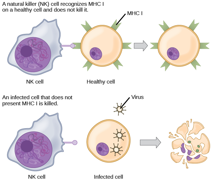
Plasma cells and CTLs are collectively called effector cells : they represent differentiated versions of their naïve counterparts, and they are involved in bringing about the immune defense of killing pathogens and infected host cells.

Mucosal surfaces and immune tolerance

The innate and adaptive immune responses discussed thus far comprise the systemic immune system (affecting the whole body), which is distinct from the mucosal immune system. Mucosal immunity is formed by mucosa-associated lymphoid tissue, which functions independently of the systemic immune system, and which has its own innate and adaptive components. Mucosa-associated lymphoid tissue (MALT) , illustrated in [link] , is a collection of lymphatic tissue that combines with epithelial tissue lining the mucosa throughout the body. This tissue functions as the immune barrier and response in areas of the body with direct contact to the external environment. The systemic and mucosal immune systems use many of the same cell types. Foreign particles that make their way to MALT are taken up by absorptive epithelial cells called M cells and delivered to APCs located directly below the mucosal tissue. M cells function in the transport described, and are located in the Peyer’s patch, a lymphoid nodule. APCs of the mucosal immune system are primarily dendritic cells, with B cells and macrophages having minor roles. Processed antigens displayed on APCs are detected by T cells in the MALT and at various mucosal induction sites, such as the tonsils, adenoids, appendix, or the mesenteric lymph nodes of the intestine. Activated T cells then migrate through the lymphatic system and into the circulatory system to mucosal sites of infection.

The intestine is lined with epithelial cells with hair-like cilia extending into the intestinal lumen. M cells are sandwiched between these epithelial cells, in bump-like projections in the intestinal lining. The M cells are shaped like an upside-down U, with the U forming a pocket on the interior surface. Antigens are taken up from the intestinal lumen by the M cells, and excreted into this U-shaped pocket. Dendritic cells in the pocket ingest the antigen, then migrate to an area below of the intestinal lining called a Peyer’s patch. The dendritic cells, T cells and B cells aggregate to form clumps of cells called organized lymphoid follicles. There, some T cells interact with antigen associated with MHC II on the surface of the dendritic cells. Some B cells are activated by free antigen. Some antigen-presenting dendritic cells enter the lymphatic system, where more B and T cells are activated in the lymph nodes. The B cells and T cells return to bigger bumps in the intestinal epithelium called MALT effector sites. Antibodies are secreted into the intestinal lumen.
The topology and function of intestinal MALT is shown. Pathogens are taken up by M cells in the intestinal epithelium and excreted into a pocket formed by the inner surface of the cell. The pocket contains antigen-presenting cells such as dendritic cells, which engulf the antigens, then present them with MHC II molecules on the cell surface. The dendritic cells migrate to an underlying tissue called a Peyer’s patch. Antigen-presenting cells, T cells, and B cells aggregate within the Peyer’s patch, forming organized lymphoid follicles. There, some T cells and B cells are activated. Other antigen-loaded dendritic cells migrate through the lymphatic system where they activate B cells, T cells, and plasma cells in the lymph nodes. The activated cells then return to MALT tissue effector sites. IgA and other antibodies are secreted into the intestinal lumen.

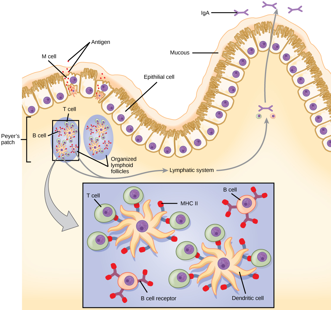
MALT is a crucial component of a functional immune system because mucosal surfaces, such as the nasal passages, are the first tissues onto which inhaled or ingested pathogens are deposited. The mucosal tissue includes the mouth, pharynx, and esophagus, and the gastrointestinal, respiratory, and urogenital tracts.

Get Jobilize Job Search Mobile App in your pocket Now!

Get it on Google Play Download on the App Store Now


Source:  OpenStax, Biology. OpenStax CNX. Feb 29, 2016 Download for free at http://cnx.org/content/col11448/1.10
Google Play and the Google Play logo are trademarks of Google Inc.

Notification Switch

Would you like to follow the 'Biology' conversation and receive update notifications?

Ask