<< Chapter < Page Chapter >> Page >
Photo (a) shows a plant with many fuzzy white hairs growing from its surface. Scanning electron micrograph (b) shows branched tree-like hairs emerging from the surface of a leaf. The trunk of each hair is about 250 microns tall. Branches are somewhat shorter. Scanning electron micrograph (c) shows many multi-pronged hairs about 100 microns long that look like sea anemones scattered across a leaf surface.
Trichomes give leaves a fuzzy appearance as in this (a) sundew ( Drosera sp.). Leaf trichomes include (b) branched trichomes on the leaf of Arabidopsis lyrata and (c) multibranched trichomes on a mature Quercus marilandica leaf. (credit a: John Freeland; credit b, c: modification of work by Robert R. Wise; scale-bar data from Matt Russell)

Below the epidermis of dicot leaves are layers of cells known as the mesophyll, or “middle leaf.” The mesophyll of most leaves typically contains two arrangements of parenchyma cells: the palisade parenchyma and spongy parenchyma ( [link] ). The palisade parenchyma (also called the palisade mesophyll) has column-shaped, tightly packed cells, and may be present in one, two, or three layers. Below the palisade parenchyma are loosely arranged cells of an irregular shape. These are the cells of the spongy parenchyma (or spongy mesophyll). The air space found between the spongy parenchyma cells allows gaseous exchange between the leaf and the outside atmosphere through the stomata. In aquatic plants, the intercellular spaces in the spongy parenchyma help the leaf float. Both layers of the mesophyll contain many chloroplasts. Guard cells are the only epidermal cells to contain chloroplasts.
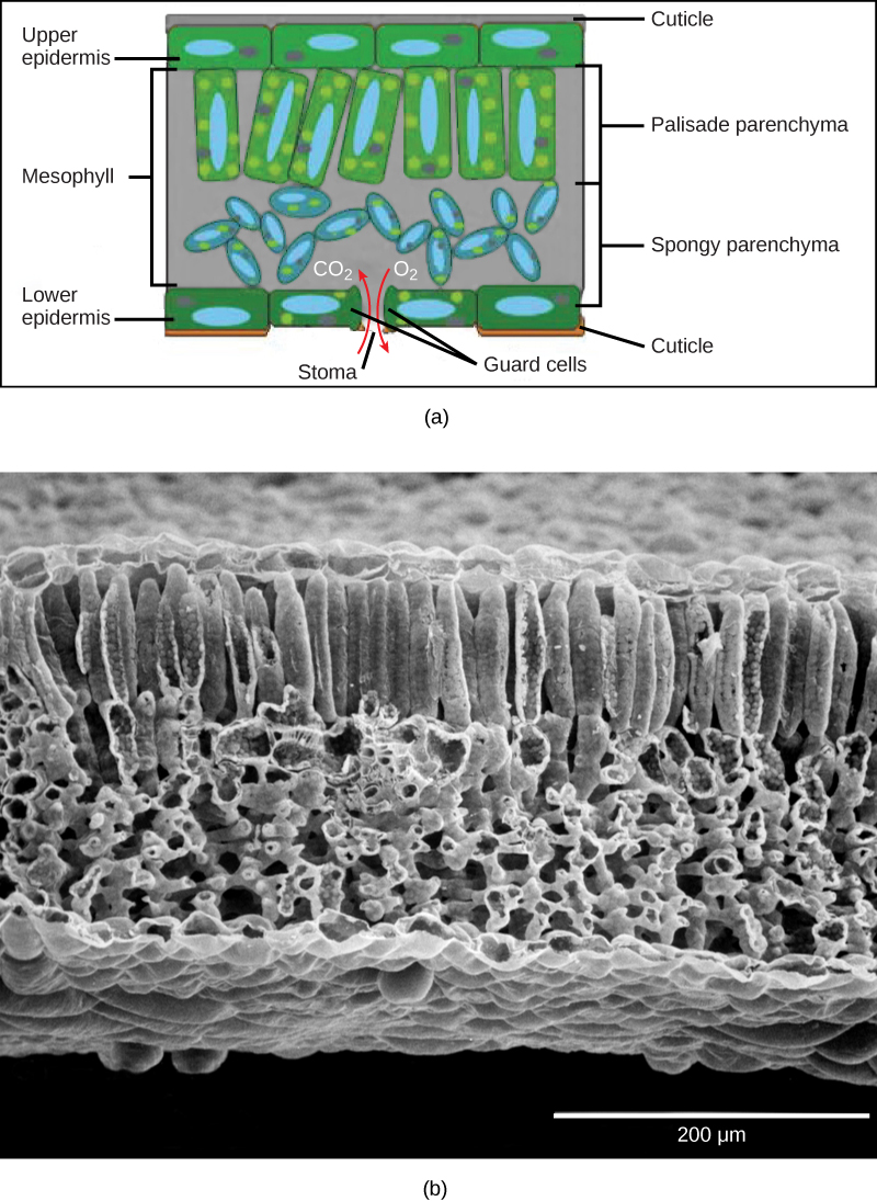
 Part A is a leaf cross section illustration. A flat layer of rectangular cells make up the upper and lower epidermis. A cuticle layer protects the outside of both epidermal layers. A stomatal pore in the lower epidermis allows carbon dioxide to enter and oxygen to leave. Oval guard cells surround the pore. Sandwiched between the upper and lower epidermis is the mesophyll. The upper part of the mesophyll is comprised of columnar cells called palisade parenchyma. The lower part of the mesophyll is made up of loosely packed spongy parenchyma. Part B is a scanning electron micrograph of a leaf in which all the layers described above are visible. Palisade cells are about 50 microns tall and 10 microns wide and are covered with tiny bumps, which are the chloroplasts. Spongy cells smaller and irregularly shaped. Several large bumps about 20 microns across project from the lower surface of the leaf.
In the (a) leaf drawing, the central mesophyll is sandwiched between an upper and lower epidermis. The mesophyll has two layers: an upper palisade layer comprised of tightly packed, columnar cells, and a lower spongy layer, comprised of loosely packed, irregularly shaped cells. Stomata on the leaf underside allow gas exchange. A waxy cuticle covers all aerial surfaces of land plants to minimize water loss. These leaf layers are clearly visible in the (b) scanning electron micrograph. The numerous small bumps in the palisade parenchyma cells are chloroplasts. Chloroplasts are also present in the spongy parenchyma, but are not as obvious. The bumps protruding from the lower surface of the leave are glandular trichomes, which differ in structure from the stalked trichomes in [link] . (credit b: modification of work by Robert R. Wise)

Like the stem, the leaf contains vascular bundles composed of xylem and phloem ( [link] ). The xylem consists of tracheids and vessels, which transport water and minerals to the leaves. The phloem transports the photosynthetic products from the leaf to the other parts of the plant. A single vascular bundle, no matter how large or small, always contains both xylem and phloem tissues.
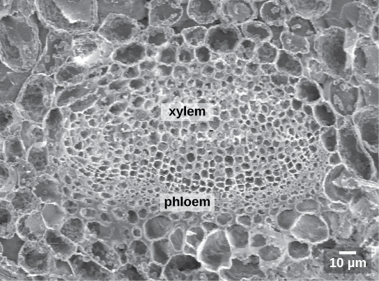
 The scanning electron micrograph shows an oval vascular bundle. Small phloem cells make up the bottom of the bundle, and larger xylem cells make up the top. The bundle is surrounded by a ring of larger cells.
This scanning electron micrograph shows xylem and phloem in the leaf vascular bundle from the lyre-leaved sand cress ( Arabidopsis lyrata) . (credit: modification of work by Robert R. Wise; scale-bar data from Matt Russell)

Leaf adaptations

Coniferous plant species that thrive in cold environments, like spruce, fir, and pine, have leaves that are reduced in size and needle-like in appearance. These needle-like leaves have sunken stomata and a smaller surface area: two attributes that aid in reducing water loss. In hot climates, plants such as cacti have leaves that are reduced to spines, which in combination with their succulent stems, help to conserve water. Many aquatic plants have leaves with wide lamina that can float on the surface of the water, and a thick waxy cuticle on the leaf surface that repels water.

Questions & Answers

what is biology
Hajah Reply
the study of living organisms and their interactions with one another and their environments
AI-Robot
what is biology
Victoria Reply
HOW CAN MAN ORGAN FUNCTION
Alfred Reply
the diagram of the digestive system
Assiatu Reply
allimentary cannel
Ogenrwot
How does twins formed
William Reply
They formed in two ways first when one sperm and one egg are splited by mitosis or two sperm and two eggs join together
Oluwatobi
what is genetics
Josephine Reply
Genetics is the study of heredity
Misack
how does twins formed?
Misack
What is manual
Hassan Reply
discuss biological phenomenon and provide pieces of evidence to show that it was responsible for the formation of eukaryotic organelles
Joseph Reply
what is biology
Yousuf Reply
the study of living organisms and their interactions with one another and their environment.
Wine
discuss the biological phenomenon and provide pieces of evidence to show that it was responsible for the formation of eukaryotic organelles in an essay form
Joseph Reply
what is the blood cells
Shaker Reply
list any five characteristics of the blood cells
Shaker
lack electricity and its more savely than electronic microscope because its naturally by using of light
Abdullahi Reply
advantage of electronic microscope is easily and clearly while disadvantage is dangerous because its electronic. advantage of light microscope is savely and naturally by sun while disadvantage is not easily,means its not sharp and not clear
Abdullahi
cell theory state that every organisms composed of one or more cell,cell is the basic unit of life
Abdullahi
is like gone fail us
DENG
cells is the basic structure and functions of all living things
Ramadan
What is classification
ISCONT Reply
is organisms that are similar into groups called tara
Yamosa
in what situation (s) would be the use of a scanning electron microscope be ideal and why?
Kenna Reply
A scanning electron microscope (SEM) is ideal for situations requiring high-resolution imaging of surfaces. It is commonly used in materials science, biology, and geology to examine the topography and composition of samples at a nanoscale level. SEM is particularly useful for studying fine details,
Hilary
cell is the building block of life.
Condoleezza Reply

Get Jobilize Job Search Mobile App in your pocket Now!

Get it on Google Play Download on the App Store Now


Source:  OpenStax, Biology. OpenStax CNX. Feb 29, 2016 Download for free at http://cnx.org/content/col11448/1.10
Google Play and the Google Play logo are trademarks of Google Inc.

Notification Switch

Would you like to follow the 'Biology' conversation and receive update notifications?

Ask