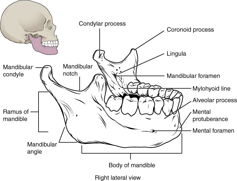
Important landmarks for the mandible include the following:
Alveolar process of the mandible —This is the upper border of the mandibular body and serves to anchor the lower teeth.
Mental protuberance —The forward projection from the inferior margin of the anterior mandible that forms the chin (mental = “chin”).
Mental foramen —The opening located on each side of the anterior-lateral mandible, which is the exit site for a sensory nerve that supplies the chin.
Mylohyoid line —This bony ridge extends along the inner aspect of the mandibular body (see
[link] ). The muscle that forms the floor of the oral cavity attaches to the mylohyoid lines on both sides of the mandible.
Mandibular foramen —This opening is located on the medial side of the ramus of the mandible. The opening leads into a tunnel that runs down the length of the mandibular body. The sensory nerve and blood vessels that supply the lower teeth enter the mandibular foramen and then follow this tunnel. Thus, to numb the lower teeth prior to dental work, the dentist must inject anesthesia into the lateral wall of the oral cavity at a point prior to where this sensory nerve enters the mandibular foramen.
Lingula —This small flap of bone is named for its shape (lingula = “little tongue”). It is located immediately next to the mandibular foramen, on the medial side of the ramus. A ligament that anchors the mandible during opening and closing of the mouth extends down from the base of the skull and attaches to the lingula.
Isolated mandible
The mandible is the only moveable bone of the skull.
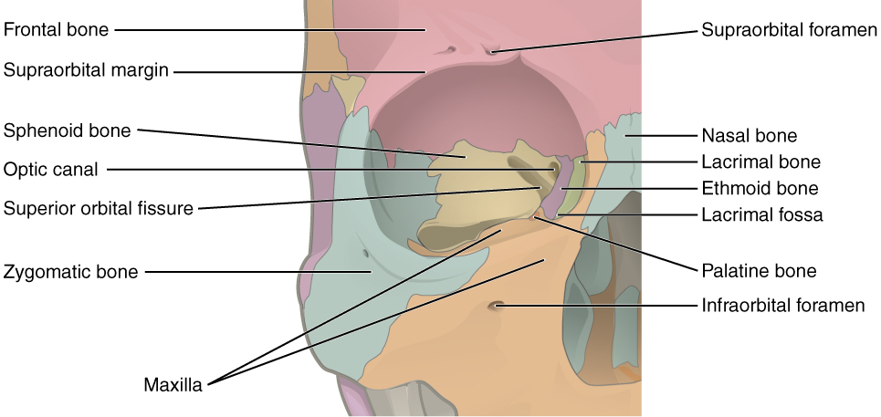
The orbit
The orbit is the bony socket that houses the eyeball and contains the muscles that move the eyeball or open the upper eyelid. Each orbit is cone-shaped, with a narrow posterior region that widens toward the large anterior opening. To help protect the eye, the bony margins of the anterior opening are thickened and somewhat constricted. The medial walls of the two orbits are parallel to each other but each lateral wall diverges away from the midline at a 45° angle. This divergence provides greater lateral peripheral vision.
The walls of each orbit include contributions from seven skull bones (
[link] ). The frontal bone forms the roof and the zygomatic bone forms the lateral wall and lateral floor. The medial floor is primarily formed by the maxilla, with a small contribution from the palatine bone. The ethmoid bone and lacrimal bone make up much of the medial wall and the sphenoid bone forms the posterior orbit.
At the posterior apex of the orbit is the opening of the
optic canal , which allows for passage of the optic nerve from the retina to the brain. Lateral to this is the elongated and irregularly shaped superior orbital fissure, which provides passage for the artery that supplies the eyeball, sensory nerves, and the nerves that supply the muscles involved in eye movements.
Bones of the orbit
Seven skull bones contribute to the walls of the orbit. Opening into the posterior orbit from the cranial cavity are the optic canal and superior orbital fissure.
the study of living organisms and their interactions with one another and their environment.
Wine
discuss the biological phenomenon and provide pieces of evidence to show that it was responsible for the formation of eukaryotic organelles in an essay form
advantage of electronic microscope is easily and clearly while disadvantage is dangerous because its electronic. advantage of light microscope is savely and naturally by sun while disadvantage is not easily,means its not sharp and not clear
Abdullahi
cell theory state that every organisms composed of one or more cell,cell is the basic unit of life
Abdullahi
is like gone fail us
DENG
cells is the basic structure and functions of all living things
A scanning electron microscope (SEM) is ideal for situations requiring high-resolution imaging of surfaces. It is commonly used in materials science, biology, and geology to examine the topography and composition of samples at a nanoscale level. SEM is particularly useful for studying fine details,