<< Chapter < Page Chapter >> Page >
Connective Tissue Examples
Connective tissue proper Supportive connective tissue Fluid connective tissue
Loose connective tissue
  • Areolar
  • Adipose
  • Reticular
Cartilage
  • Hyaline
  • Fibrocartilage
  • Elastic
Blood
Dense connective tissue
  • Regular elastic
  • Irregular elastic
Bones
  • Compact bone
  • Cancellous bone
Lymph

Connective tissue proper

Fibroblasts are present in all connective tissue proper ( [link] ). Fibrocytes, adipocytes, and mesenchymal cells are fixed cells, which means they remain within the connective tissue. Other cells move in and out of the connective tissue in response to chemical signals. Macrophages, mast cells, lymphocytes, plasma cells, and phagocytic cells are found in connective tissue proper but are actually part of the immune system protecting the body.

Connective tissue proper

The left image shows a diagram of connective tissue. As a whole, the connective tissue appears somewhat disorganized, with fibers and cells mixed together heterogeneously. There are many open spaces between the embedded elements, suggesting that the connective tissue is somewhat loosely packed. The thickest fibers are collagen fibers; the thinner fibers are elastic fibers. Both the collagen fibers and the elastic fibers crisscross randomly throughout the tissue. In addition, a net of reticular fibers appear in the upper part of the diagram. Two yellow and oval shaped adipocytes are embedded below the reticular fiber net, with a small dark nucleus squeezed into one corner of the cell. A mesenchymal cell is next to one of the adipocytes. The cell is rectangular and has four projections stemming from each corner of the cell. The projections appear to attach to the nearby collagen fibers. A fibroblast is located at the center of the diagram. The fibroblast appears similar to the mesenchymal cell, except that it is larger and has more projections. Finally, a white macrophage is in the lower right of the diagram. The macrophage is a white, oval shaped disc with a prominent nucleus. The right diagram is a micrograph of connective tissue. The tissue is mostly stained pink, however, the thick collagen fibers crisscrossing the tissue are white. Five adipocytes also appear white, except for their cell membrane and nucleus, which stained dark. A mesenchymal cell occupies the space between two adipocytes. It stains a very deep purple, but its shape is unclear in the micrograph. A fibrocyte is also visible as an oval shaped cell with a deep purple nucleus.
Fibroblasts produce this fibrous tissue. Connective tissue proper includes the fixed cells fibrocytes, adipocytes, and mesenchymal cells. LM × 400. (Micrograph provided by the Regents of University of Michigan Medical School © 2012)

Cell types

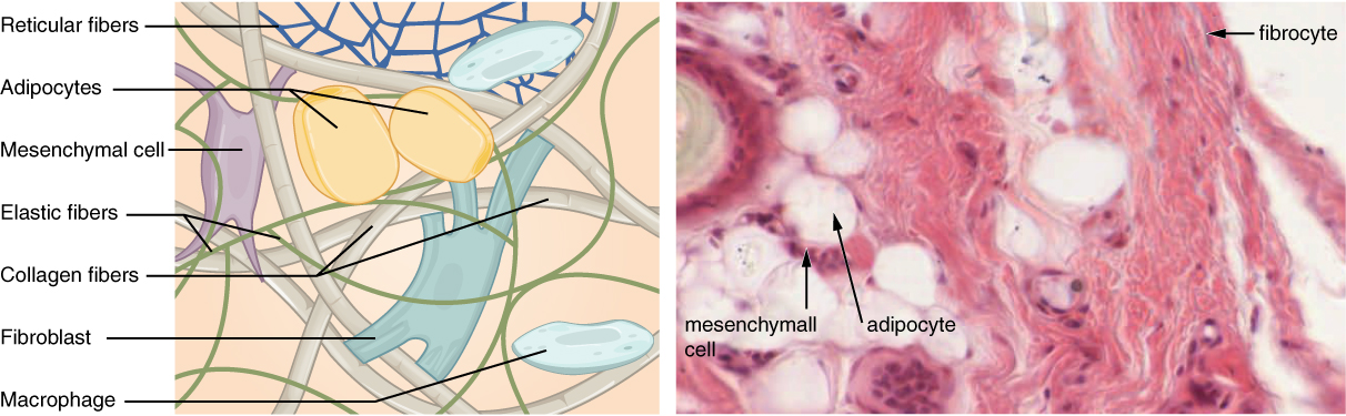
The most abundant cell in connective tissue proper is the fibroblast    . Polysaccharides and proteins secreted by fibroblasts combine with extra-cellular fluids to produce a viscous ground substance that, with embedded fibrous proteins, forms the extra-cellular matrix. As you might expect, a fibrocyte    , a less active form of fibroblast, is the second most common cell type in connective tissue proper.

Adipocytes are cells that store lipids as droplets that fill most of the cytoplasm. There are two basic types of adipocytes: white and brown. The brown adipocytes store lipids as many droplets, and have high metabolic activity. In contrast, white fat adipocytes store lipids as a single large drop and are metabolically less active. Their effectiveness at storing large amounts of fat is witnessed in obese individuals. The number and type of adipocytes depends on the tissue and location, and vary among individuals in the population.

The mesenchymal cell    is a multipotent adult stem cell. These cells can differentiate into any type of connective tissue cells needed for repair and healing of damaged tissue.

The macrophage cell is a large cell derived from a monocyte, a type of blood cell, which enters the connective tissue matrix from the blood vessels. The macrophage cells are an essential component of the immune system, which is the body’s defense against potential pathogens and degraded host cells. When stimulated, macrophages release cytokines, small proteins that act as chemical messengers. Cytokines recruit other cells of the immune system to infected sites and stimulate their activities. Roaming, or free, macrophages move rapidly by amoeboid movement, engulfing infectious agents and cellular debris. In contrast, fixed macrophages are permanent residents of their tissues.

The mast cell, found in connective tissue proper, has many cytoplasmic granules. These granules contain the chemical signals histamine and heparin. When irritated or damaged, mast cells release histamine, an inflammatory mediator, which causes vasodilation and increased blood flow at a site of injury or infection, along with itching, swelling, and redness you recognize as an allergic response. Like blood cells, mast cells are derived from hematopoietic stem cells and are part of the immune system.

Questions & Answers

what is phylogeny
Odigie Reply
evolutionary history and relationship of an organism or group of organisms
AI-Robot
ok
Deng
what is biology
Hajah Reply
the study of living organisms and their interactions with one another and their environments
AI-Robot
what is biology
Victoria Reply
HOW CAN MAN ORGAN FUNCTION
Alfred Reply
the diagram of the digestive system
Assiatu Reply
allimentary cannel
Ogenrwot
How does twins formed
William Reply
They formed in two ways first when one sperm and one egg are splited by mitosis or two sperm and two eggs join together
Oluwatobi
what is genetics
Josephine Reply
Genetics is the study of heredity
Misack
how does twins formed?
Misack
What is manual
Hassan Reply
discuss biological phenomenon and provide pieces of evidence to show that it was responsible for the formation of eukaryotic organelles
Joseph Reply
what is biology
Yousuf Reply
the study of living organisms and their interactions with one another and their environment.
Wine
discuss the biological phenomenon and provide pieces of evidence to show that it was responsible for the formation of eukaryotic organelles in an essay form
Joseph Reply
what is the blood cells
Shaker Reply
list any five characteristics of the blood cells
Shaker
lack electricity and its more savely than electronic microscope because its naturally by using of light
Abdullahi Reply
advantage of electronic microscope is easily and clearly while disadvantage is dangerous because its electronic. advantage of light microscope is savely and naturally by sun while disadvantage is not easily,means its not sharp and not clear
Abdullahi
cell theory state that every organisms composed of one or more cell,cell is the basic unit of life
Abdullahi
is like gone fail us
DENG
cells is the basic structure and functions of all living things
Ramadan
What is classification
ISCONT Reply
is organisms that are similar into groups called tara
Yamosa
in what situation (s) would be the use of a scanning electron microscope be ideal and why?
Kenna Reply
A scanning electron microscope (SEM) is ideal for situations requiring high-resolution imaging of surfaces. It is commonly used in materials science, biology, and geology to examine the topography and composition of samples at a nanoscale level. SEM is particularly useful for studying fine details,
Hilary
Got questions? Join the online conversation and get instant answers!
Jobilize.com Reply

Get Jobilize Job Search Mobile App in your pocket Now!

Get it on Google Play Download on the App Store Now


Source:  OpenStax, Anatomy & Physiology. OpenStax CNX. Feb 04, 2016 Download for free at http://legacy.cnx.org/content/col11496/1.8
Google Play and the Google Play logo are trademarks of Google Inc.

Notification Switch

Would you like to follow the 'Anatomy & Physiology' conversation and receive update notifications?

Ask